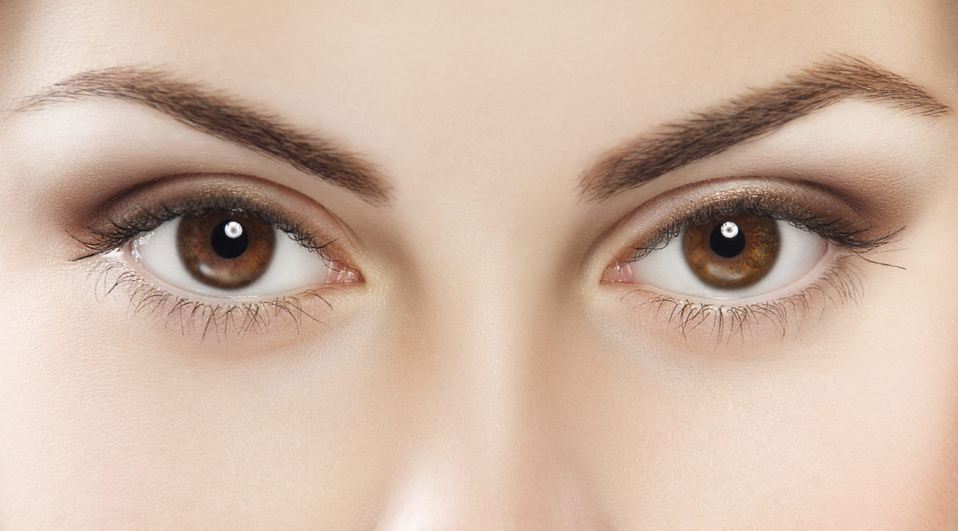
The eye is a sensory organ that obtains light around us and converts it into electrical nerve impulses which are then interpreted as a visual image thus providing sight. There are different eye conditions, including common eye ailments and defects such as long or short-sightedness, cataracts, conjunctivitis, and dry eyes. However, there are many other intricate and complicated eye conditions that can cause more problems to your health. You stand to gain a lot by getting yourself familiar with some common eye ailments and defects. The goal is to detect minor eye conditions early on, thus preventing them from becoming a major health issue.
Top Related Article: The Importance of Eye Care
This article explains in detail, some different types of eye conditions, including common eye ailments and defects, as well as well as rare ones. It also explains what symptoms are associated with each and how they can be treated.
12 Types of Eye Conditions
1. LONG AND SHORT-SIGHTEDNESS
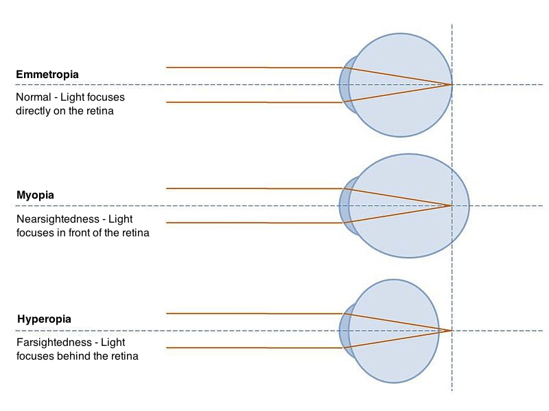
Long-sightedness (Hyperopia) and Short-sightedness (Myopia) are common eye conditions that occur when light rays do not focus on the retina properly.
Hyperopia
Hyperopia, also known as long-sightedness, is an eye condition that occurs when the eyeball is shorter than it’s supposed to be–i.e. the anteroposterior diameter of the eye is short, or the cornea is not curved enough. This causes light to focus too far back behind the eye. Consequently, the image of a close object is formed behind the retina, causing it to appear blurred. However, this eye condition doesn’t affect distant vision because the light rays reflected from them strike the retina as parallel rays, so it remains clear.
Symptoms of Hyperopia
- Close up objects appear blurry
- Reading becomes difficult especially when the book is at arm’s length
- Headaches and migraine may occur as a result of over straining the eyeballs
Causes of Hyperopia
- Age: Babies and children generally tend to be naturally long-sighted. This, however, is very slight and their eyes lengthen a little as they grow up. Also, Presbyopia or age-associated long-sightedness occurs when the eye becomes very stiff and loses it focusing power.
- Genetic Inheritance: No specific long-sightedness gene has been identified by research, but it is generally thought to be an inherited eye condition.
- Other Underlying Conditions: Long-sightedness can be caused by other conditions such as diabetes, orbital tumors, small eye syndrome or microphthalmia (a condition in which one or both eyes are abnormally small, with anatomical abnormalities) and foveal hypoplasia (a condition characterized by underdevelopment of the macula, an area on the retina responsible for clear and detailed vision).
Myopia
Myopia also known as short-sightedness is an eye condition that occurs when the anteroposterior diameter of the eyeball is longer than normal, or the cornea is too steeply curved. This allows light rays to be focused in front of the retina causing distant objects to appear blurred.
Symptoms of Myopia
- Distant objects appear blurry
- Children might have difficulty seeing the whiteboard in their classroom.
- Frowning and squinting
- Headaches may occur as a result of overstraining the eyeballs
Causes of Myopia
- Age: Myopia usually occurs from early childhood up to 21 years, the risk of one getting it reduces with age. It might not be easily detected in children because they may be able to see closer things and do their homework without any difficulty, but they might not be able to see distant objects so well.
- Genetic Inheritance: It has been discovered from research that there’s a 30% chance of a child developing myopia if one of the parents have it and a 55% chance if both parents have it.
- Environmental factors: Straining the eyes by doing close-range tasks for a long time can lead to myopia. These tasks involve staring at a computer or a book for too long.
Treatment of Long and Short-sightedness
There are many treatment methods to treat this eye condition. They include:
- Spectacles: This is the cheapest and most common form of treating a refractive problem. They cause no harm to the eye and offer some form of protection in case of an accident. Convex lenses can be used to correct long-sightedness while Concave lenses can be used to correct short-sightedness. Wearing correct lens ensures light rays fall on the retina enabling you to focus correctly.
- Contact Lenses: Contact lenses can also be used to correct this eye problem, they carry regular expense and a number of risk factors but they are generally preferred because they offer the freedom of participating in rigorous activities. They also improve personal appearance because they are almost invisible.
Contact lenses can be worn daily (daily disposable lenses), disinfected for re-use (weekly disposable lenses), or worn for a longer period of time. (monthly disposable lenses). There other types of contact lenses such as Rigid Gas Permeable, Multifocal / Bifocal, and Hard contact lenses. Your optometrist will advise you on the lens type that will best suit you.
- Surgical Treatment: Though traditional surgical methods are available for treating this eye condition, the most reliable surgical treatment method involves laser surgery. The shape of the cornea is changed by removing some tissues from the edge to compensate for the reflective problem and to further increase the focusing power of the eyes. Another advantage of the laser surgery method is that it reduces the risk of getting another eye infection or damaging the eye.
The major types of laser surgery include
-
-
- Laser in situ keratectomy (LASIK)
- Photorefractive keratectomy (PRK)
- Laser epithelial keratomileusis (LASEK)
-
Your surgeon will advise you on the best laser surgery procedure to be used for you. Laser surgery methods also carry their risk complications which occur rarely. It can lead to
- Ectasia: a condition in which the cornea becomes very thin and leads to reduction or loss of vision.
- Correction error: a situation whereby the surgeon miscalculates the amount of tissue needed to be removed from the cornea
- Epithelial ingrowth: This occurs when the flap cut into the cornea starts growing into the main part of the cornea. This may be corrected with further surgery.
2. PRESBYOPIA
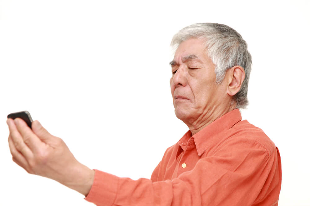
Presbyopia is a naturally occurring eye condition associated with the ageing of the eye. It is a more common eye problem in humans as they get older. The lens of the eye continues to grow throughout one’s lifetime causing the inner fibers to become tightly compacted. As a result of this, the focusing power of the eyes reduces with age. By the time a person reaches forty, these changes start to become more noticeable. The person starts moving objects (like a book or a newspaper) further away from the eyes so they can see it clearly.
Top Article: What is Eye Strain?
The eye lens of a child is still very elastic, so it changes shape easily enabling them to focus on both nearby and distant objects. The lens stiffens as a person grows thus reducing the lens flexibility.
Symptoms of Presbyopia
- Difficulty in near vision and reading small prints
- Asthenopic Symptoms: They occur due to fatigue of the ciliary muscles caused by doing a close-range work
- Headache occurring after completing close-range work
Causes of Presbyopia
- Age-related changes in lens: This might include a progressive increase in size and hardness of lens or decrease in the elasticity of the lens as one grows older.
- Other causes: Presbyopia is an age-related eye condition but there are factors that can cause premature presbyopia, they include chronic glaucoma, premature hardness (sclerosis) of the crystalline lens, uncorrected hyperopia and diabetes.
- Drugs: Some drugs have been discovered to cause premature presbyopia, examples of such drugs include diuretics, antidepressants, and antihistamines
Treatment of Presbyopia
Thankfully, there are ways to treat this type of eye condition.
- Glasses: This the most common ways to correct presbyopia. Depending on the peculiarity of each person’s case, a varifocal or bifocal corrective lens can be used to correct presbyopia
- Contact Lenses: For those who do not like glasses, contact lenses are an option to correct the loss of focusing power caused by Presbyopia.
- A common example is the Lasik Blended Vision (monovision) Treatment in which one eye is corrected for distance and the other eye is corrected for close-range.
- Surgery: New surgical procedures have been developed over the years to correct presbyopia. Implantation of an Intraocular lens (IOL) is a good example of a surgical procedure used to treat presbyopia, it involves changing the eye’s optical power by turning the corneas into multifocal lenses.
3. CONJUNCTIVITIS
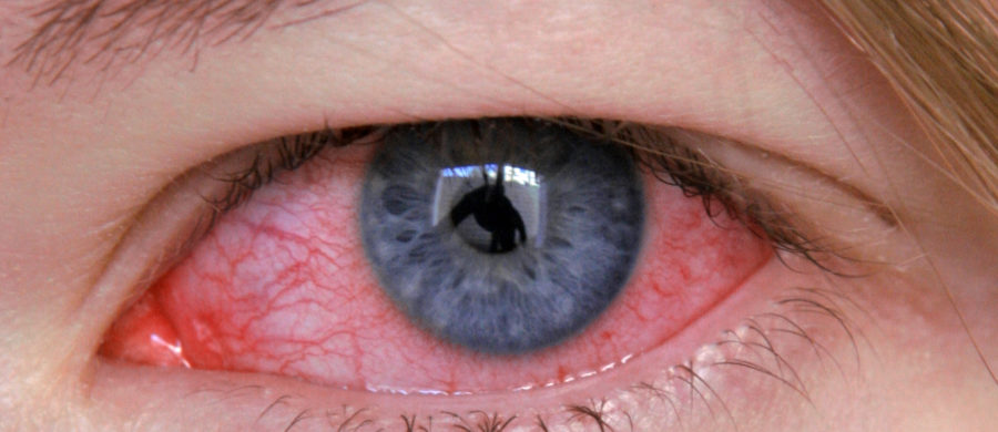
Conjunctivitis or pinkeye is a common eye problem in humans. This eye condition is caused by the inflammation or irritation of the conjunctiva (lining of the eye) caused by allergies, bacterial or viral infection.
Major types of conjunctivitis include infective conjunctivitis, allergic conjunctivitis, and irritative conjunctivitis. Although, there are other forms of conjunctivitis which occur in rare cases such as Keratoconjunctivitis and Traumatic conjunctivitis.
Infective conjunctivitis
This is the most common eye problem in humans related to conjunctivitis. It is the inflammation of the conjunctiva caused by microorganisms such as a virus, bacteria or a sexually transmitted infection (STI) like Chlamydia. If a mother has Chlamydia, she can pass it to her baby during delivery which can lead to conjunctivitis in the child.
Allergic conjunctivitis
The conjunctiva is very sensitive to allergens, up to ten times more than the skin. Allergic conjunctivitis is the inflammation of the conjunctiva caused by allergic or hypersensitivity reactions to an allergen. This reaction may occur immediately (humoral) or might be delayed (cellular).
Irritative conjunctivitis
This occurs when an irritant such as chlorine, dust or an eyelash enters into the eye and makes it sore for some time. This type of conjunctivitis goes away once the irritant settles. It is imperative for a person not rub their eyes because it can worsen the situation and the debris can scratch the eye.
Symptoms of Conjunctivitis
- Sensation of a foreign body in the eye due to inflammation of vessels.
- Mucopurulent discharge from the eyes; the conjunctiva contains a lot of cells that produce mucus and tiny glands, irritation of these glands can lead to water discharge from the eyes.
- Sticking together of lid margins: this is more noticeable when you wake up in the morning. The eyelids feel like they are stuck together because of sticky lumps on the lashes produced formed by the mucus produced by the infection.
- Red-eye: This occurs when the blood vessels in the conjunctiva are widened.
Treatment of Conjunctivitis
Like other eye conditions on this list, there are treatment options available for conjunctivitis.
- Antibiotics: Antibiotics are not always necessary because most forms of conjunctivitis clear on their own after a few days or weeks. When the conjunctivitis is severe or it has lasted for a couple of weeks without clearing, antibiotics may be prescribed for such person. Common types of antibiotics prescribed are Chloramphenicol, Ciprofloxacin and Fusidic acid.
- Hygiene and self-care:
-
- Wash your hands regularly to stop the infection from spreading to other people.
- Clean sticky substances from your eyes when you wake up in the morning using a damp cotton wool.
- Contact lenses should be removed until all signs and symptoms of conjunctivitis are gone
- Use lubricant eye drops. They can be purchased over-the-counter (OTC) at your local drug store. They may help ease any soreness and stickiness in your eyes.
- Steroid drops
Viral conjunctivitis will not respond to antibiotic drops. Instead, it is treated by the use of artificial tears, over-the-counter painkillers, cold compresses and regular cleaning of the eye. Extreme cases of viral conjunctivitis can be treated with steroid drops.
4. ASTIGMATISM
Astigmatism is a common eye condition that occurs when the surface of the cornea has an irregular curve and is more shaped like a rugby ball. Consequently, light rays entering the eye cannot converge to focus directly on the retina forming a blurred image. Between 30 and 60% of adults in Europe and Asia have astigmatism. It is also an eye condition caused by refractive error also like long-sightedness and short-sightedness.
It is possible to have some degree of astigmatism in one or both eyes while just one eye has eye hyperopia or myopia.
Related: Eye Strain Causes
There are two main types of Astigmatism: regular and irregular Astigmatism. Irregular astigmatism is caused when there’s an irregular change of refractive power in different meridian often noticed when there’s a corneal scar or ‘scattering’ in the eye’s crystalline lens. Regular astigmatism is caused when the refractive power changes uniformly from one meridian to another.
Symptoms of Astigmatism
- Astigmatism affects both close and distant vision, objects at all distances seem blurred
- Eye strain and headaches may occur as a result of focusing on one thing for too long.
- Objects may appear proportionately elongated depending on the type and degree of your astigmatism.
Causes
Causes of this common eye condition include:
- Light is not properly focused on the retina when it hits an irregularly curved cornea. This focus blurs the image, resulting in blurred vision.
- Hereditary factors: Some people are born with this eye condition which is why astigmatism is generally believed to be hereditary.
- Other factors: Astigmatism could occur as a result of–
- Keratoconus – a rare eye condition in which the cornea gets weaker, thinner and tends to change shape.
- Changes to the cornea caused by a previous surgery.
- Injury to the cornea
Treatment
- Glasses: corrective lenses can be can be tolerated up to a maximum difference of 4 Diopters after Diplopia occurs which is a situation whereby a person sees 2 images instead of one.
- Contact lenses: this form of treatment is usually advised for people with a chronic case of astigmatism. Toric lenses can be prescribed for regular astigmatism while rigid gas-permeable contact lens may be used in more severe cases.
- Laser eye surgery: This treatment procedure involves the shaping of the cornea to correct the refractive problem. Laser eye surgery procedures such as Laser in situ keratectomy (LASIK) and Photorefractive keratectomy (PRK) are very successful in treating astigmatism.
5. DRY EYE
Dry eye is one of the most common eye ailments and defects. This eye condition causes the eyes to not make enough tears, or they evaporate the tears too quickly. Tears consist of water, oil, mucus and antibodies, all of which help to lubricate the eye, stabilize vision, wash away debris, and protect it against infection. When there are not enough tears on the surface and beneath the lids of the eye, the eye starts to itch, as well as feel dry and irritated.
Symptoms of Dry Eye
- The eyes constantly feel dry, gritty or tired
- Vision starts to get slightly blurry
- Red eyes
- Eyelids may stick together after waking up in the morning
Causes of Dry Eye
- Age and gender: Dry eye is a more common eye condition in older generations. That’s because your eyes produce fewer tears as you age. Research has shown that around 75% of all people aged over 65 are affected by dry eyes.
- Gender is another factor, with women more likely to be affected than men. This is as a result of hormonal changes in women associated with lactation, pregnancy, menstruation and menopause.
- Side effects from taking certain medications: Taking medications such as decongestants, blood pressure medication, antihistamines or beta-blockers, oral contraceptives, and antidepressants may have side effects leading to dry eyes.
- Focusing on visual tasks such as using a computer for a long period of time causes less blinking than normal. While using a computer you may blink around 7 times per minute, rather than the normal rate of around 22 times per minute. This causes increased evaporation of tears leaving the eye dry.
- Other medical conditions can lead to dry eyes such as rheumatoid arthritis, Sjogren’s Syndrome, diabetes, asthma, thyroid disease, blepharitis or eyelid conditions.
Treatment of Dry Eye
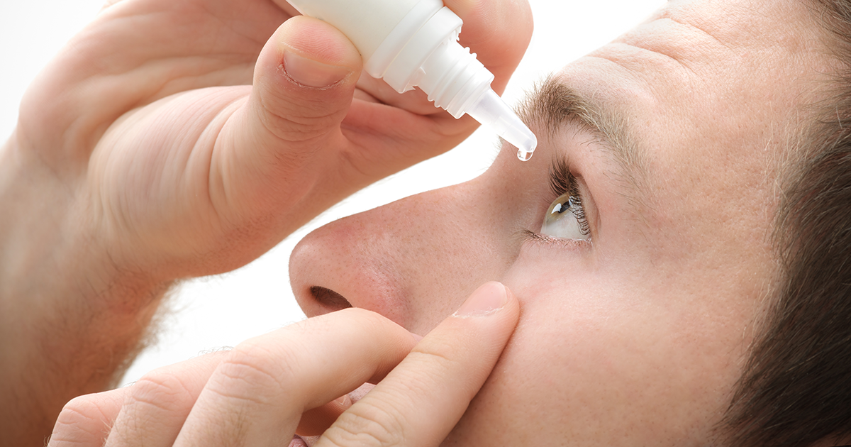
- Tear substitutes: This is the most common form of dry eye treatment. Tear substitutes include eye drops, gels or ointments. Most dry eye conditions can be treated using products that contain tear substitutes, a liquid designed to imitate the properties of eye tears. These products are can be purchased over the counter from a pharmacy without a prescription. No single product can work for everyone, so you may need to try a few products before figuring out which one works best for you.
You might need to use eye drops frequently if you have a chronic dry eye. Some eye drops contain preservatives to keep bacteria from growing inside it. If you have a severe dry eye or you wear soft contact lenses, it is better to get an eye drop that is preservative-free so it does not further irritate your eye.
Thicker substances like ointment can be used if your eyes get dry while you sleep. It should be the last thing you use at night because it can cause blurred vision. Eye ointments that contain paraffin should not be used if you wear contact lenses.
- Surgery: surgery is a last resort form of treatment if your chronic dry eye just won’t go away no matter what you try. There are two common types of surgery used to correct dry eyes:
- Punctual occlusion: Punctual occlusion involves using plugs to seal the punctum, the duct that drains tears from your eye. These small plugs stop tears from draining, as a result, your eyes remain moist all the time. Temporary plugs made of silicone are designed to dissolve over time, they are first tried to determine whether the operation has a positive effect on your dry eye condition or not. Permanent plugs or non-dissolving punctual plugs are then used if the temporary plugs work well.
Your doctor might also opt out for a procedure known as cautery in which a special tool is used to burn the exit duct shut. Tear plugs make your eyes feel better because your eyes are more adapted to natural tears rather than artificial ones.
-
- Salivary gland auto transplantation: This is a surgical procedure used for treating severe dry eye conditions. It involves removing some glands that produce saliva from the lower lip for placement or grafting into the side of the eyes.
- Lipiflow Treatment also known as thermal pulsation involves the use of a medical device that uses heat and pressure to clear blocked glands on the eyelids. These glands are responsible for producing oil in the tears to keep them from evaporating, thereby keeping the eye moist.
- Testosterone cream: Dry eye condition caused by lack of testosterone in the oil glands can be treated by applying a testosterone cream to the eyelid. Also, drugs such as Cyclosporine (Cequa, Restasis) and Lifitegrast (Xiidra) can help kick-start and boost tear production.
- Nutrition and hydration can also go a long way in the treatment of dry eye conditions. Natural supplements such as Fish oil, flax seed oil, and vitamin E together with Omega-3 supplements can be added to your diet to help treat dry eye. Examples of products that combine some or all of these supplements are TheraTears Nutrition capsule and Lovaza Omega-3 acid ethyl esters capsule.
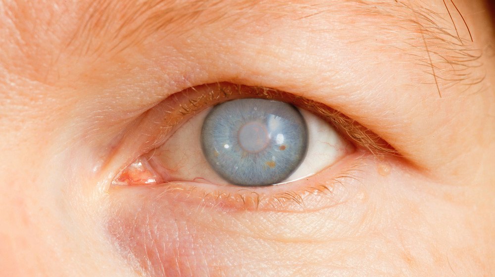
6. CATARACTS
Cataract is a common type of eye condition. It reduces the quality of the image formed on the retina as a result of clouding the normally clear lens of the eye. It is a common eye problem in humans. The lens of the eye is naturally crystalline enabling us to focus on objects at varying distances. As we grow older, the eye lenses develop cataracts and reduction in vision starts to occur because the eye can’t focus light properly on the retina. Cataracts is perhaps one of the most common types of eye conditions in the world, with over 90% of people experiencing it at some point in their lifetime.
Symptoms of Cataracts
- Vision is blurry, cloudy or foggy
- Changes in the way color is seen, it may appear faded or washed out
- Poor night vision
- Double vision in the affected eye
- Problems with driving at night
- Difficulty in reading or seeing objects at a distance (Nearsightedness)
- Double vision in the affected eye
- Eyeglasses or contact lenses not working well
Causes of Cataracts
- Age-related Cataracts: This kind of cataract forms as one grows older. It affects both men and women equally and its commonly found in people who are over 65 years.
- Congenital Cataracts: This is a term used to describe when babies are born with cataracts. Thus, can be due to poor womb development, injury or infection.
- Cataracts can be caused by other medical conditions such as diabetes, excessive use of medication, injury to the eye, exposure to toxic substances including excessive Ultra Violet Rays from the sun, and other eye conditions such as Uveitis.
Treatment of Cataracts
Cataracts cannot be prevented, you can only make some lifestyle changes to slow down its formation. Smoking, excessive alcohol intake, direct exposure to excessive sunlight, obesity and high blood pressure are all modifiable factors that can aid the formation of cataracts.
Cataracts are treated by having a surgery to remove the foggy lens in the eye, this can be replaced with an artificial plastic lens. This artificial lens is also knowns as intraocular lens (IOL).
The procedure is carried out in a day and you may or may not need to sleep over in the hospital. More than 95% of people who have undergone this procedure say they can now see better and have relief from this common eye condition. Cataracts cannot be treated with laser surgery; the most common cataract operation is phacoemulsification, also known as phaco extracapsular extraction. Different types of intraocular lenses used in cataract surgery include a standard monofocal intraocular lens, a multifocal intraocular lens or a toric intraocular lens.
- Monofocal intraocular lens is a fixed lens designed to offer a fixed focus at one distance (usually far distances). The patient might need to get reading glasses to see near objects even if the person didn’t wear glasses before the surgery.
- Multifocal intraocular lens is designed to deliver improved vision over a range of distances both far and near. The patient might not need to wear glasses after surgery. However, Multifocal intraocular lenses are not right for every patient, some patients may experience glare when driving at night while others might have difficulty adjusting to their new vision.
- Toric intraocular lenses are designed for individuals with astigmatism, they offer a broader range of vision. Toric lens can correct both cataract and astigmatism, but the patient might be required to use reading glasses.
7. GLAUCOMA
Glaucoma is one of the most common types of eye conditions. It occurs when the drainage tubes (trabecular meshwork) within the eye become slightly blocked preventing the eye fluid (aqueous humor) from draining properly. Intraocular pressure begins to build up and reaches a point where it becomes powerful enough to eventually damage the optic nerve connecting the brain to the eye. Glaucoma may affect one or both eyes and can cause permanent blindness if not properly treated. In most people, glaucoma shows no early symptoms or pain, you have to see the eye doctor regularly to diagnose and treat glaucoma before it becomes worse. If you have a family history of glaucoma, you should probably a get an eye checkup every 1-2 years.
There are four major types of glaucoma namely chronic open-angle glaucoma, primary open angle glaucoma (POAG), secondary glaucoma and congenital glaucoma
- Chronic open-angle glaucoma: this type of glaucoma is very common eye problem in humans and it develops very slowly.
- Primary angle-closure glaucoma: this is a rare type of glaucoma, it can occur very slowly or suddenly develop pressure build-up on the eye.
- Secondary glaucoma: this type of glaucoma occurs as a result of a previous eye injury or another eye condition, such as uveitis (inflammation of the middle layer of the eye).
- Congenital glaucoma: this occurs rarely but can be very severe. It is a situation whereby a child develops glaucoma at birth or shortly after birth.
Symptoms of Glaucoma
- In cases of Chronic open-angle glaucoma, most people don’t notice any symptoms until late in the disease, that is why glaucoma is often referred to as the “sneak thief of vision.”
The symptoms are barely noticeable because the outer field of vision (peripheral vision) is the first part of the eye to be affected before it slowly works inwards towards the center of the eye.
- For acute angle-closure glaucoma, the symptoms are severe, they may include:
- Redness in the eye
- Intense eye pain
- Seeing halos or ‘rainbow-like’ rings around lights
- Misty and narrowed vision
- Quick progression of reduction of vision in one or both
- For secondary glaucoma, the symptoms of glaucoma can be confused with the symptoms of the other condition. An example is uveitis which often causes pain in the eyes and headaches. However, secondary glaucoma may still cause seeing halos around light and misty vision
- Congenital glaucoma: it may be very difficult to detect glaucoma in children. However, some symptoms that are more commonly displayed include:
- Sensitivity to light (photophobia)
- Watery eyes and misty vision
- Large eyes due to the pressure in them
- Squint, an eye condition that occurs when one of the eyes turn inwards, outwards or upwards, while the other eye looks straight.
Visit your optometrist if you notice any of these symptoms.
Causes of Glaucoma
- Age: Glaucoma is more likely to affect older people.
- Short-sightedness (myopia): People with myopia are more likely to develop chronic open-angle glaucoma.
- Ethnic origin: Africans or Afro-Caribbeans are more likely to develop chronic open-angle glaucoma while Asians or people from Asian origin are more likely to develop acute angle-closure glaucoma.
- Hereditary factor: if you have a family history of glaucoma or you have a close relative that has it, you are more likely to develop the eye condition yourself. Regular eye tests should be done to know the condition of your eyes.
- Other underlying medical conditions such as diabetes have also been highly associated with glaucoma.
Treatment of Glaucoma
Treatment may vary slightly between different eye units and the population being treated.
- Eye drops: This is usually the first line of treatment. Some eye drops can increase the outflow of the aqueous humor fluid (Prostaglandin analogues) while some reduce the intraocular pressure on the eye or reduce the formation of the fluid (beta-blockers). Side effects of using eyedrops may include enlarged blood vessels in the white part of the eye, thick and dark eyelashes, eye pain and irritation, redness, blurred vision, and itchy eyes.
- Laser treatment.
- Laser trabeculoplasty involves the use of high energy beams of light to open up the blocked trabecular meshwork (drainage tubes) within the eye.
- Iridotomy: It’s a procedure that involves making a tiny hole in the iris to let fluid flow more freely.
- Cyclophotocoagulation: This procedure involves treating areas of the middle layer of the eye in order to reduce fluid production.
- Microsurgery: The most common type is trabeculectomy in which a new channel is created to drain the fluid and ease pressure on the eye. There are also other types of glaucoma surgery such as viscocanalostomy and aqueous shunt implant.
Viscocanalostomy is an operation that involves removing part of the sclera (the white outer covering of the eyeball), to filter the fluid out of the eye and into the body. Aqueous shunt implant is an operation that involves placing a tube device into the eye to assist in the drainage of the fluid.
Your surgeon can discuss each procedure and the associated risks to help you make the best decision to treat your eye conditions.
8. AMBLYOPIA (LAZY EYE)
Amblyopia, also known as lazy eye, is an early childhood eye condition in which the eye and the brain do not work together, resulting in decreased vision. It sometimes occurs in both eyes but on very rare occasions. Lazy eye affects about 2-3% of all children and can be treated if caught early on. The eye condition can be very difficult to treat after it has developed which is usually about age 7-8.
Symptoms of Lazy Eye
If a child has a lazy eye, they will not receive clear images through the faulty eye, causing the brain to receive an unclear image. The good eye will often try to make up for the affected eye making the eye become lazy. Children might not be able to notice if they have a lazy eye or not until they go through an eye test.
Major symptoms of a lazy eye may include blurred or double vision, droopy eyelid, noticeable squint (an eye condition that occurs when one of the eyes turns inwards, outwards or upwards, while the other eye looks straight), and cataracts.
Causes of Lazy Eye
There are many different eye disorders that lead to a lazy eye, they include
-
- Strabismic amblyopia: This is the most common cause of lazy eye; a squint is manifested in one because the eyes look in different directions. It can be caused by hereditary factors, long or short-sightedness, a viral disease or an injury.
- Anisometropic amblyopia: This occurs when the light is not focused properly on the retina due to short-sightedness, long-sightedness or astigmatism. In this case, the brain ignores signals from the affected eye which results in the eye becoming lazy.
- Ametropic amblyopia: It occurs when a child has large refractive errors causing an unclear image to form on the retina. It leads to a reduction in the visual quality of both eyes.
- Stimulus deprivation amblyopia: This is the rarest and most severe form of lazy eye. It happens when one eye or both eyes do not see properly and therefore become lazy.
Treatment of Lazy Eye
- Using a patch: The most common method of treating a lazy eye is to force the brain to start using the weak eye. Underlying problems in that eye such as nearsightedness, farsightedness, or astigmatism are first treated. A patch is then given to the child to wear over the strong eye in order to force the weak eye to work. It might take weeks or months, but the child’s vision should eventually get better over time.
- Atropine Eye drops: They dilate the pupil of the good eye causing near vision to blur. This encourages the use of the lazy eye. Side effects that may occur from using these eye drops include eye irritation, flushing (reddening) of the skin, and headaches.
- Eye surgery: Surgery may be needed if the lazy eye is caused by another problem, such as a cataract or a squint. This form of treatment can also be used in conjunction with the patch treatment. Surgery can also be used to realign the lazy eye, thus improving binocular vision.
- Vision therapy: This form of treatment is sometimes used to help a child’s vision develop. Selected exercises and games that require the child to use the affected eye are carried out.
9. KERATOCONUS
Keratoconus is a progressive eye disorder that occurs when the normally round cornea becomes thin and conical shaped. This abnormal shape prevents light rays from being focused properly on the retina, leading to distortion of vision and increased sensitivity to glare and light. Other symptoms include double vision, myopia, blurred vision, and astigmatism, usually affecting both eyes. Most people that experience this eye condition begin to develop it in their late teens or early 20s.
Symptoms of Keratoconus
Visual quality is affected once the cornea begins to change its shape. Because the cornea is responsible for most of the eye’s focusing, a change in shape may result in blurred and distorted vision, astigmatism, inability to see in dim light, double vision, nearsightedness, sensitivity to light, or vision loss. In addition, it could result in a rim of discoloration around the front of the eye known as the Fleischer ring.
Related: Eye Strain Symptoms
Glasses and contact lenses become less effective and uncomfortable as keratoconus progresses because the cornea is now conical.
Causes of Keratoconus
Though some studies have shown that keratoconus is associated with asthma, eczema and excessive eye rubbing, it is not known exactly what causes keratoconus. However, it is thought to be an inherited condition.
Treatment of Keratoconus
Keratoconus can be treated in different ways; an example is the use of corneal implant which involves placing small pieces of plastic in the front part of the eye to correct the shape of the cornea. Corrective and contact lenses may also be placed over the eyes to improve and correct vision. Surgery can also be done to replace the cornea with a donated one.
An advanced method of treatment is corneal collagen cross-linking which involves the application of riboflavin solution to the eye which is activated by illumination with UV-A light. This causes new bonds to form across adjacent collagen strands and makes the cornea stronger.
10. MACULAR DEGENERATION
Macular degeneration is an eye condition caused by the deterioration of the macular (i.e. the small central part of the retina) leading to impaired vision. It is often referred to as age-related macular degeneration (AMD) because it is an eye condition that develops as a person grows older. Macular degeneration cannot be cured but it can be treated with vitamins and medications, laser therapy, and vision aids. Although it never blinds a person totally, it can be a source of significant visual disability.
Symptoms of Macular Degeneration
Macular degeneration is not a painful condition and may not be discovered until the symptoms become really severe. The major symptom of macular degeneration is blurring of central vision. The central vision is responsible for the perception of fine details and colors. Other symptoms associated with Macular degeneration include:
- Loss of visual acuity: this is the loss of the ability to notice little details or read across small distances.
- Loss of contrast sensitivity: this is the loss of the ability to see less well-defined objects, such as faces.
Causes of Macular Degeneration
- Dry age-related macular degeneration: This form of macular degeneration is characterized by the presence of yellow deposits, called drusen, in the macula. As they grow in size and increase in number, distortion of vision starts to occur. In the atrophic form of dry macular degeneration, patients may have blind spots at the center of their vision. Severe case of macular degeneration can lead to loss of central vision.
- Wet age-related macular degeneration: This form of macular degeneration is characterized by the growth of abnormal blood vessels underneath the macula. The blood vessels start to leak blood and fluid into the retina, causing distortion of vision. Permanent loss of central vision can occur when the abnormal blood vessels eventually form a scar.
- Risk factors: There are many factors that can increase the risk of getting macular degeneration, they include age, gender, genetic factors, smoking, exposure to too much sunlight and excessive intake of alcohol
Treatment of Macular Degeneration
There is currently no cure for macular degeneration, but there are many treatment options that can slow down the progression of the disease or prevent severe vision loss. These include:
- Anti-angiogenesis drugs. These drugs block from the abnormal vessels within the eye that cause wet macular degeneration. This treatment can help to regain vision that was lost. Example of these drugs include: Aflibercept, Macugen, Eyelea, and Lucentis.
- Laser therapy. This is a procedure in which high-energy laser lights are used to destroy actively growing abnormal blood vessels that occur as a result of macular degeneration.
- For possible prevention of macular degeneration: vitamins C and E, beta-carotene, zinc, and copper can decrease the risk of patients losing their vision due to macular degeneration.
11. UVEITIS
Uveitis is an eye condition caused by the inflammation of the middle layer of the eye known as the uvea or uveal tract. The uvea is made up of the iris, the choroid (layer of tissue supporting the retina) and the ciliary body (ring of muscle behind the iris)
There are many types of uveitis, they are classified based on which part of the eye is affected. They include:
- Anterior Uveitis: This is the most common type of uveitis caused by the inflammation of the iris and/or the ciliary body. It accounts for 3 out of every 4 cases of uveitis.
- Intermediate uveitis: This type of uveitis affects the area behind the ciliary body and the retina. It occurs more in children and teenagers and accounts for 1 out of every 5 cases of uveitis.
- Posterior Uveitis. This type of uveitis affects the area behind the eye, the choroid, and the retina. Most of the time it is related to an underlying autoimmune condition, such as rheumatoid arthritis.
Symptoms
The symptoms of uveitis include:
- Painful red eye
- Blurred or cloudy vision
- Floaters: which refers to shadows that move across the field of vision
- Discoloration of the iris
- Loss of peripheral vision caused by posterior uveitis
Causes
The most common cause of uveitis is by inflammation, this has helped to identify two major causes of uveitis namely infectious uveitis and non-infectious uveitis. Infectious Uveitis is caused by inflammation in response to a real infection while Noninfectious uveitis usually occurs in people who have an underlying autoimmune condition. The immune system malfunctions and triggers the process of inflammation even though no infection is present.
Other causes of uveitis include Lyme disease, tuberculosis, injury to the eye, and syphilis in rare cases.
Treatment
- Viruses are the most common cause of infectious uveitis which can be treated with antiviral medications or antibiotics.
- Corticosteroid eye drops: This is the major type of medication used to treat non-infectious uveitis. Recommended dose can be as frequent as an eyedrop per hour or as intermittent as an eye drop once every two days.
- Corticosteroid injections: When the symptoms fail to respond to the eye drops, you may need to have a corticosteroid injection. The injection is administered on the side of the eye so as not to damage it.
- Oral corticosteroids which may come in the form of tablets or capsules are recommended if other treatments don’t work or if it’s interfering with your daily activities,
- Immunosuppressants: These are used as a last resort in case a patient fails to respond to other treatments previously discussed. Immunosuppressants are used to control the immune system and to disrupt the process of inflammation.
12. BLEPHARITIS
Blepharitis is an eye condition caused by the chronic inflammation of the eyelids. It is a chronic condition which means it can resurface after it has been treated. It can lead to red swollen eyelids and also itchy and soreness in the eyes. In severe cases, the eyelashes may fall out which may lead to other eye conditions such as styes and small ulcers.
Symptoms
Symptoms of blepharitis differ depending on the causative agent. Major symptoms may include:
- Itchy, sore, red eyelids
- Sticky eyelids especially when waking up in the morning
- Crusty, or oily and greasy eyelids
- Increased sensitivity to light (photophobia)
- Abnormal eyelash growth and/or loss of eyelashes
- Swollen eyelid margins (the edges of your eyelids)
Causes
There are two major types of blepharitis namely Anterior blepharitis and Posterior blepharitis.
- Anterior blepharitis: It is majorly caused by bacterial infection, usually a staphylococcal infection. It can also occur as a complication of seborrheic dermatitis (a skin condition that causes your skin to become inflamed or flaky). Blepharitis that is caused by seborrheic dermatitis is often referred to as seborrheic blepharitis.
- Posterior blepharitis: It is caused when there is a problem with the Meibomian glands found on the rim of your eyelids. The Meibomian glands are responsible for producing the oily substance present in tears.
Treatment
- Eye hygiene: Cleaning the eyelids can go a long way in the treatment of blepharitis. Effective eye hygiene will help reduce the severity and frequency of the symptoms caused by this eye condition.
- Topical antibiotics: This form of treatment is used when the eye condition doesn’t respond to regular cleaning. Topical antibiotics such as chloramphenicol eye ointment and fusidic acid eye drops can be used to treat blepharitis.
- Oral antibiotics: This form of treatment is administered when it is clear that a skin condition, such as rosacea, is aggravating the eye condition. Oral antibiotics may also be recommended if the eye condition doesn’t respond to other forms of treatment. Some oral antibiotics products are known to make people more sensitive to the sun. Therefore, avoid excessive exposure to sunlight while on an oral antibiotic treatment.
