Eye anatomy is fascinating—there is so much going on behind the surface of your eyes that allows them to work as they do and process the world around you. It is difficult to identify one sense as more important than another; however, many individuals might argue that vision is the most important of the five senses. It is also often reported as the mode of perception that people would fear to lose the most. Why? Being able to see is an extremely important ability that allows us to not only visualize the world but also utilize this unique sense in combination with the other senses to interact and understand the world. Imagine trying to go through your everyday routine without being able to see. How would you turn the light on? How would you know where anything is? Clearly, sight is important. In fact, more than 75% of the information we receive about the world is through vision. All in thanks to anatomical features of the eye.
The organ that allows us and the majority of animals to see is the eye. Often, the eye is compared to an anatomical version of a camera. Each eye individually gathers light and utilizes it to create a picture. Just like a camera, each eye has several parts that allow us to take this raw stimulus of light and transform it into a picture that we can interpret as vision. In this article, we will cover each different component of the eye and the role it plays in allowing us to see. Please keep in mind that the anatomy of the eye is more complex and in-depth than the discussion in this article; this article is designed to provide a comprehensive introduction to the anatomy of the eye.
Top Related Article: Vision Care
Before we begin with eye anatomy itself, it is important to first discuss the anatomy surrounding the eye. This includes the orbit, eyelids, eyelashes, and extraocular muscles.
Anatomical Features of the Eye: Orbit, Eyelids, and Eyelashes
The orbit is the bony socket in the skull in which the eye itself lies. It’s formed by multiple bones, including the cheekbone, forehead, temple, and side of the nose. It not only contains the eyeball, but it also contains the multiple muscles that move the eye, its blood vessels and nerves. The eyelids seem like they have no role, other than cosmetic, yet they serve an important purpose. They protect the eye from any foreign matter, including dust, dirt, foreign objects, and even extremely bright light that can lead to subsequent eye damage. The eyelids also spread tears and lubrication over the surface of your eye as you blink. The eyelashes serve as an additional filter and protective layer.
Extraocular (External) Muscles
What are these extraocular muscles? They are muscles that surround the outside of the eye. These extraocular, external, are attached to the eye which allows it to move in different planes of motion and rotate, allowing us to have a broad field of vision that isn’t limited by the eyeballs’ ability to move. In humans, we have a total of six extraocular muscles, and these six muscles are further divided into two classes: the rectus and oblique, both of which play slightly different roles in allowing the eye to move.
– Rectus Muscles
We will start with the rectus muscle. There are four different rectus muscles that surround the eye. The first is the superior rectus, and as indicated in its Latin roots, this muscle attaches itself to the top of the eye. Similarly, based on its placement, it allows the eye to move upward. On the opposite end of the eye is the inferior rectus, which attaches itself to the bottom of the eye. Contrastingly, it moves the eye downward. The last two rectus muscles attach themselves to each side of the eye. The lateral rectus attaches itself to the outside of the eye, attaching itself near the temple, and allows the eye to move outward. The medial rectus is the last rectus muscle, which attaches itself to the side of the eye near the nose. This muscle allows the eye to move inward towards the nose, thus playing an important role in eye anatomy.
– Oblique Muscles
The remaining two extraocular muscles are the oblique muscles. The superior oblique originates from the back of the orbit and attaches itself near the top of the eye. Its role is different to that of the superior rectus, as it rotates the eye inward along the sagittal plane (front to back). It can also move the eye downward. The inferior oblique originates near the nose, around the front of the orbit. It then wraps along the bottom of the eye, traveling out laterally, and attaches on the bottom of the eyeball. It rotates the eye outward along the sagittal plane and can move the eye upward.
Eye Anatomy – How Your Eyes Really Work
Conjunctiva
So, let’s slowly travel inward into the eye itself from these extraocular muscles. Next is the conjunctiva. This is a protective membrane that covers the surface of the eye and the inner surface of the eyelids. It is transparent and mucous-like in nature. It is made up of two segments: the bulbar and palpebral conjunctiva. The bulbar conjunctiva covers the anterior, front, portion of the sclera. It stops where the sclera and cornea meet each other, and it does not cover the cornea. The palpebral conjunctiva covers the inner portions of both eyelids on each eye. These two conjunctivae are continuous, and this is why it is impossible for anything to get caught behind your eye, such as your contact lens. Now, why do we have this layer? It keeps the front of the eye, and inner surfaces of the eyelids well lubricated. This allows this portion of the eye and eyelids to move with minimal friction and prevents irritation. The conjunctiva also acts as a protective layer for the eye.
Related: Treatment Options for Eye Conditions
Lacrimal Gland/ Tenon’s Capsule
The lacrimal gland is a small gland that produces tears, which then act as an additional lubricant for the eye. It is located under the outside of the eyebrow, contained in the orbit. The Tenon’s capsule is another layer that lies between the conjunctiva and the surface of the eye. Now let’s move into the anatomy of the eye itself.
Sclera
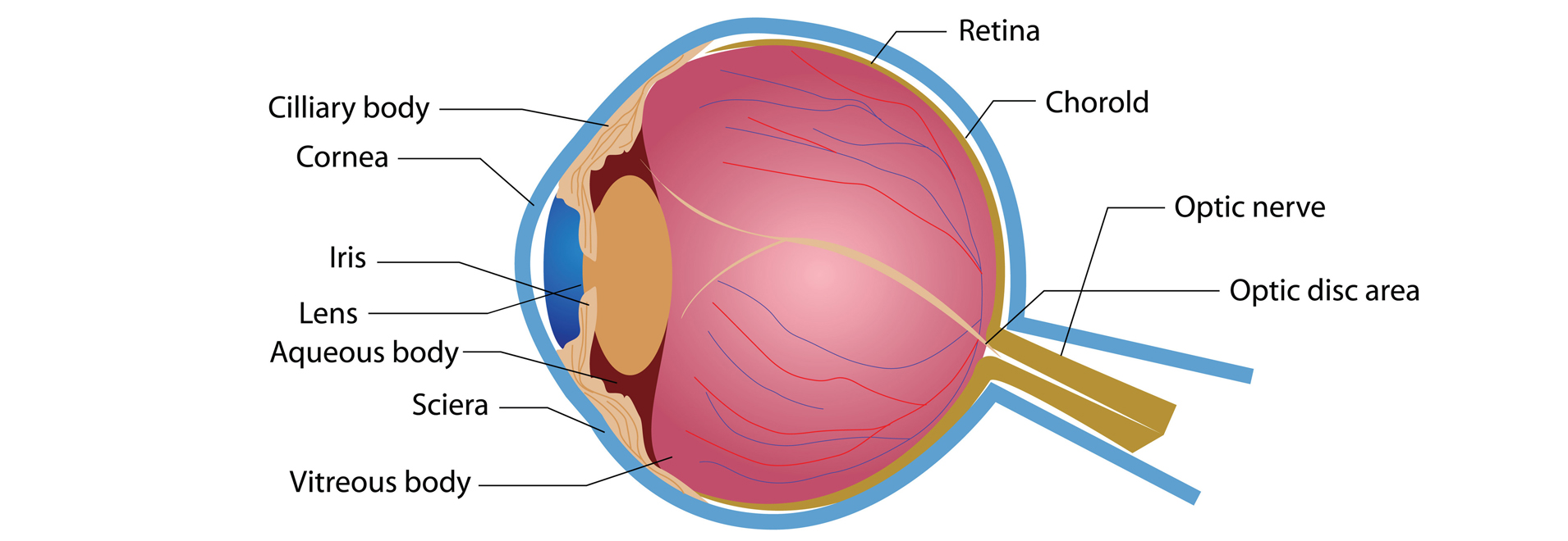
The sclera is the white layer of the eye which surrounds the cornea. It is commonly known as the “white part of the eye.” The sclera composes a large majority of the surface area of the eye – more than 80 percent of the surface area of your eye. It begins from the cornea and wraps behind all the way to the optic nerve at the back of the eye.
What is the sclera layer? It is a dense layer of connective tissue, composed of multiple connected interweaved bundles of collagen. Unlike other layers of the eye, the sclera is considered avascular, meaning it lacks blood vessels. This subsequently also means that it has a limited blood supply and compared to other layers of the eye, its metabolic activity is relatively low. The sclera plays an important role in eye health. It helps maintain the round shape of the eyeball in combination with intraocular pressure (IOP). As previously mentioned, its multiple layers through the connective network of collagen, the sclera protects the eye from serious damage. It also acts as a sturdy foundation on which the extraocular muscles can connect and control the fine motor movements of the eye.
Cornea
The cornea is the first point in the eye through light enters. It lies in front of the iris and pupil; it is the lens through which light enters the eye. Although it may look clear, it is organized and serves multiple purposes. Because it lacks blood vessels, it receives its nourishment from tears. It is made up of five different layers: the corneal epithelium, Bowman’s layer, corneal stroma, Descemet’s membrane, and corneal endothelium.
Corneal epithelium: This is the outermost layer of the cornea and comprises 10 percent of the total thickness of the cornea. Its main functions are to protect the eye from the foreign material while also acting as a surface in which oxygen and nutrients from tears can be absorbed. From this layer, oxygen and nutrients are distributed to the other layers of the cornea.
Bowman’s membrane: This is an extremely thin layer that serves as a connective layer between the corneal epithelium and stroma. It is made up of collagen.
Stroma: This is the thickest layer of the cornea, making up around 90 percent of the cornea’s thickness. This is the layer that gives the cornea its strength, elasticity, and shape. It is made up of largely water and collagen. The uniform arrangement of collagen fibers is what allows the cornea to be essentially transparent.
Descemet’s membrane: Similar to the Bowman’s layer, the Descemet’s membrane serves as another protective layer between the stroma and corneal endothelium. It is also made up of collagen fibers, although the composition of these collagen fibers differs from the stroma.
Corneal endothelium: This is the innermost layer of the cornea, and the back of this layer is bathed in a clear fluid, the aqueous humor, which occupies the space between the cornea and iris and pupil. Endothelial cells play an important in keeping the cornea clear. They pump the excess fluid out of the stroma, as it leaks slowly. Without this, the stroma would swell with fluid, losing its clear nature.
As previously mentioned, the cornea acts as the first layer in which light enters the eye, acting as a barrier for foreign materials. Yet, it also has another important role. The cornea is responsible for between 65 and 75 percent of the eye’s ability and power to focus. Light is bent, or refracted, from the cornea onto the lens, which then further focuses light onto the retina. This will be further discussed later in the article.
Intraocular Anatomy
Now, we will be discussing the internal, intraocular anatomy.
Anterior Chamber/ Posterior Chamber
This is a fluid-filled space that lies immediately behind the cornea and continues until the front of the iris. It is filled with a fluid called the aqueous humor that provides nourishment to both the cornea and lens. Similar to the anterior chamber, there is another fluid-filled layer called the posterior chamber, which is behind the iris but in front of the lens. It plays an identical role to that of the anterior chamber.
Uvea
Next is the uvea. The uvea is the middle layer of the eyeball, made up of three different components: the iris, ciliary body, and choroid. The uvea is the pigmented layer of the eye.
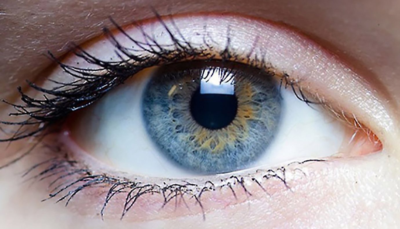
Iris: The iris is a thin, circular structure that surrounds the pupil. This layer is also responsible for your eye color, which is based on the amount of pigment in this layer. The iris also acts to control the size of the pupil. One muscle in the iris constricts in bright light and another muscle in the iris dilates in the dark.
Ciliary body: This second part of the uvea holds the lens in place. It surrounds the iris but cannot be seen as it is behind the sclera. It also secretes the aqueous humor, which was previously mentioned as the fluid that fills the space between the cornea and the iris and lens.
Choroid: This is the innermost layer of the uvea, which has the important role of providing nourishment to the retina. Because of this role, it contains numerous tiny blood vessels; this layer is located between the sclera and the retina.
Pupil
Ironically, the pupil, a crucial part of the eye, is an open space. The pupil serves as an opening, in the center of the iris, through which light can enter the eye, so it can be further focused on the retina and allow us to visualize the world. Unlike the iris, the pupil appears black in color, and this is because the light that passes through the pupil is absorbed and not reflected back by the retina. The iris essentially surrounds the pupil. It works hand in hand with the iris, and together, they control how much light enters the eye. Using the previous camera analogy, the pupil acts as the aperture and the iris acts as the diaphragm which controls the size of the aperture. The size of the pupil is also controlled by the muscles of the iris.
Lens
Directly behind the pupil is the lens. As previously mentioned, it is held in place by the ciliary bodies. The lens acts to further bend light, allowing it to better focus light onto the retina.
Vitreous Body
The vitreous cavity makes up a large portion of the back of the eye. Similar to the anterior and posterior chambers, it is filled with a fluid called the vitreous humor. This fluid nourishes the inner structures of the eye. It is the portion of the inner eye behind the lens leading up to the retina.
Retina – Cells
The retina is the important part of eye anatomy that allows us to essentially see. It is a layer of nerve cells lining the back portion of the eye, in which the raw light stimulus is converted into signals that the brain can interpret, resulting in our sense of vision. It is composed of multiple layers of millions of specialized cells packed closely together, photoreceptor cells. Humans have two different types of photoreceptor cells that play different roles in our vision: rods and cones. Rod photoreceptors are responsible for detecting motion and provided black and white vision. Additionally, they are able to function fairly well in low light. They are located throughout the retina and outnumber cone cells tremendously.
Cone photoreceptors are responsible for color, performing best in medium and bright light. They are also responsible for central vision. Because of the varying frequencies in wavelength that result in different colors, humans have three types of cone photoreceptors that vary in specific sensitivity to these different wavelengths.
Top Related Article: The Importance of Eye Care
Unlike rods, cones are concentrated in a small area in the middle of the retina called the macula. There is another central small portion of the macula called the fovea, where there are only cone photoreceptors. Maximum visual acuity and color vision occurs at the fovea. It is estimated that each retina has around 120 million rods and 6 million cone photoreceptors. As previously mentioned, each of these photoreceptors is a cell that contains an axon.
There is another class of photoreceptors in the retina called retinal ganglion cells. These cells are much less common, but they still play an important role. They can be stimulated by light even when all other rod and cone photoreceptors are blocked. They contain a pigment called melanopsin, and changes in this pigment by light occur in non-image forming processes, such as synchronization of circadian rhythms.
There are also numerous supporting neural cells in the retina, including bipolar cells, ganglion cells, horizontal cells, and amacrine cells. Bipolar cells are located entirely within the retina, and their role is to connect photoreceptor cells to ganglion cells They are postsynaptic to rods and cones, meaning they help continue transmitting the signal further down. Ganglion cells have dendrites that essentially connect them with bipolar cells, and they communicate with each other through synapses. The axons of ganglion cells make up the nerve fiber layer that then becomes the optic nerve fibers in the brain.
Horizontal cells are laterally interconnecting neurons that connect bipolar cells with each other. They also play a role in allowing our eyes to adjust to different intensities of light. Amacrine cells connect bipolar and ganglion cells. It acts as a connecting cell that can affect and modify the output of bipolar cells, and similar to horizontal cells, they are oriented laterally, whereas bipolar and ganglion cells act vertically.
There are other supporting glial cells in the retina. These include Muller cells, astrocytes, and microglial cells. These glial cells are located throughout the axons of the ganglion cells and the optic nerve, serving various roles in mediating various neural processes.
There is an additional layer in the retina, called the Retinal Pigment Epithelium, which is a layer of cells located deep in the retina. Its role is to help maintain the function of photoreceptor cells. It does so through the processing of Vitamin A, absorbing light, and transporting nutrients to and from the photoreceptor cells.
Anatomic Layers of the Retina
Now that we’ve discussed all of the cells that make up the retina, let’s discuss the anatomic layers of the retina. The retina is made up for ten different layers, and we will discuss them from the innermost layer, closest to the vitreous, and moving outwards toward the sclera.
The inner limiting membrane is the layer between the vitreous body and retina. Next is the nerve fiber layer, which contains the optic nerve fibers – ganglion cell axon fibers. The ganglion cell layer contains the nuclei of the ganglion cells and also contains the retinal ganglion cells. The inner plexiform layer contains the synapses between the dendrites of the ganglion and amacrine cells and the axons of the bipolar cells. The inner nuclear layer houses the nuclei of the horizontal, bipolar, and amacrine cells.
The outer plexiform layer has the axons of the rod and cone cells and the dendrites of the horizontal and bipolar cells. The outer nuclear layer houses the rod and cone cell bodies. Lastly, the outer limiting membrane separates the cell nuclei of the photoreceptors from their inner portions. The inner and outer segments of photoreceptors cells, both rods, and cones are located in the rod and cone layer. Lastly, the pigment epithelium is the outermost layer of the retina.
The Eye to the Brain

The final components of eye anatomy now focus on leaving the eye through the optic nerve and traveling towards the brain. The optic nerve, as its name implies, is the nerve that connects each eye to the brain. It travels from each eye towards the brain, and both of these optic nerves meet at a point called the optic chiasm. This is where there is a visual crossover which is why the right side of the brain controls the left visual field whereas the left side of the brain controls the right visual field.
Following the transmission of the signal from the optic chiasm, the signal continues into the brain where it is processed in an area called the visual cortex. This is where vision occurs, allowing us to understand visual information, such as color, composition, size, shape, and relation to other objects in space.
So how does vision occur?
Now that we’ve gotten a lot of the gross eye anatomy out of the way, it is time to discuss the process of how light is converted into neural signals, and ultimately better understand the process of how we see. There are four processes underlying the greater process of converting light into neural signals: photoreception, transmission to bipolar cells through synapses, transmission to ganglion cells, and transmission along the optic nerve.
Photoreception
Light travels from its source through all of the previously mentioned layers of the eye, reaching the inner layers of the retina where the photoreceptors lie. The light must past through and around the ganglion cells before reaching the rods and cones. Rods and cones contain photopigment on their outer segments, which take individual photons of light. This serves as the raw stimulus used to start the process of neuronal signaling.
The inner portion of photoreceptor cells contains the axon terminal, where neurotransmitters are released, further transmitting the signal to the nearby bipolar cells. As previously mentioned, rods work better in dim light settings, providing black and white lights. Contrastingly, cones are much more effective in well-lit situations, such as daytime, and provide the perception of color.
The outer segments of photoreceptor cells transduce light and their subsequent signals are sent out. This signal continues and reaches the bipolar and horizontal cells. The horizontal cells act to help in signal processing and the bipolar cells continue transmitting this signal to the ganglion cells. The ganglion cells then send their axons back to the optic nerve which continues until the optic chiasm where the optic nerves of both eyes meet. There is a crossover, and the signal continues traveling down the optic tract until the light signal is processed in the primary visual cortex of the brain.
As previously mentioned, this article was written to provide you with a comprehensive overview of eye anatomy. Although we understand that this was a lot of information and some of it may have been overwhelming, we would like to note that there is a lot of material that was not fully discussed in depth. Understanding the human body and the processes that occur simultaneously, that we often take for granted, requires a tremendous amount of research, and we still do not understand it all. Yet, we hope that this article can act as a solid foundation for understanding the anatomy of the eye and a basic understanding of how that anatomy leads to our sense of vision.
