Having a basic understanding of human eye anatomy is key to understanding how technology is impacting your eyes. From a young age, many of us are told to look at people in their eyes. A person’s eyes can show if they are scared, excited, or telling a lie. We have even placed safety precautions into law that protect our eyes. The human eye is so important to daily functions. You wear sunglasses when it is bright or turn on a light when it is dark. We want our eyes to be protected and to function properly when we need them. We never really think about the anatomical structure of our eye. You may think that only the outside of them, or the cornea, gets hurt. In this article, we will explore the anatomy of the human eye. With a basic understanding of human eye anatomy, we will explore what technology has done to help our eyes, but also what harm technology can do to our eyes.
Top Related Article: Symptoms and Signs of Eye Health
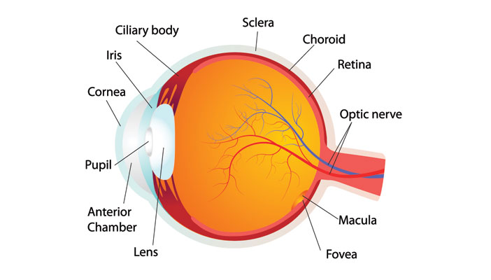
The anatomy of the human eye consists of twelve major parts. From the outside, most of what people know about the anatomy of the eye is clearly visible. There are the whites of eye, the pupil, the iris, and some veins. The eye seems so simple that many forget the complex anatomical structure that exists behind the eyelids. To understand the role that technology plays in our eyesight, one must first understand human eyeball anatomy.
Human Eye Anatomy
Cornea
The cornea is the transparent front part of the human eye. Although the cornea is clear and appears to be weak, it is very strong and durable. The cornea covers the iris, pupil, and the anterior chamber. One of its purposes is to protect the other parts of the eye from germs, dust, and other harmful materials. In conjunction with the anterior chamber and the lens, the cornea refracts light. The cornea accounts for nearly two-thirds of the eye’s total optical power. The cornea also acts as a filter as it is responsible for blocking some of the most harmful ultraviolent sunlight. Without the cornea, the lens would be in jeopardy of being damaged by UV light.
Iris
The iris is the pigmented curtain near the front of the eye. It is located between the cornea and the lens. It is shaped like a doughnut and the missing part in the middle is known as the pupil. The iris consists of two thin strips of muscles. These muscles are responsible for the dilation and contraction of the pupil. The amount of pigment in the iris determines the eye color of the person. A lack of pigment means that a person will have blue eyes. If a person has more pigment, they will have dark-colored eyes.
The purpose of the iris is to control the amount of light the retina receives. The iris will dilate the pupil in low light and will constrict it in bright lights. The iris will guarantee that the retina receives the optimal amount of light without a person needing to make a conscious decision. An interesting fact about the iris is that no two peoples’ iris is the same. They are much like the fingerprints of the eye.
Ciliary Body
The ciliary body is the part of the eye that connects the iris to the choroid. The choroid will be explained later in this article. The ciliary body consists of the ciliary muscle, the ciliary process, and the ciliary ring. The ciliary muscle is responsible for altering the curvature of the lens. This alteration allows the eye to focus on objects that are near. The ciliary process is a series of ligaments that keep the lens at the optimal position in the eye. The ciliary ring joins the ciliary body to the choroid. Lastly, the ciliary body is part of the eye’s vascular system and it helps the eye receive nourishment.
Pupil
The pupil appears as a black circle in the center of the iris. The pupil is actually a void in the iris where nothing exists. The muscles that make up the iris control the size of the pupil so that the appropriate amount of light will pass through it and reach the retina. The reason the pupil is black is that light is not reflected back from the retina. Any light that the pupil allows to enter the eye is absorbed by the retina. Any time the pupil appears to be anything other than completely black, there is some type of medical issue affecting the lens.
When observing a person going from dark to light, the pupil constricts to a small size very quickly. This is known as the pupillary reflex. The pupil will constrict much further than it needs to and then adjust as the retina responds to the ideal amount of light. Much like the iris, the pupil varies in size between people. It is interesting that the pupil is considered part of the eye when it really is just a void.
Sclera
The sclera is the largest part of the eye. It is the white part of the eye that surrounds the cornea. The sclera is 80% of the eye’s surface. The sclera has a few very important functions. It is responsible for maintaining the shape of the eyeball. It also protects the eye against external trauma. Another feature of the sclera is that it is the attachment point for the extraocular muscles that control the movement of the eye. The next part of the eye helps protect the sclera and cornea.
Conjunctiva
The conjunctiva is a mucous membrane that covers the front of the eye and the inside of the eyelids. The conjunctiva serves the eye in several different ways. It keeps the eye moist and lubricated. Along with lubricating the eye, it lubricates the inner eyelids to prevent friction. The conjunctiva protects the eye from dust, debris, and infection-causing microorganisms. The conjunctiva produces a substance that makes up part of a tear. This substance plays an integral role in preventing dry eye syndrome. The conjunctiva also acts as a vessel to transports nutrients to the rest of the eye and the eyelids. The next part of the eye we will cover has everything to do with sight and nothing to do with protecting the eye itself.
Lens
The lens is the clear, elastic part of the eye behind the iris. It is the second part of the eye that helps focus light and images on the retina. The lens is responsible for up to one-third of the eye’s focusing power. As stated above, the lens is attached to the ciliary muscles. These muscles can change the lens to focus light onto the retina in order to form a clear image. The lens is very flexible, but that flexibility decreases with age. As we age, the lens can no longer flex as much as it did in our youth. That is why many older individuals require some time of corrective eye glasses.
Macula
The macula is an oval-shaped pigmented area near the center of the retina. The macula is responsible for high-resolution, color vision that is possible in optimal lighting. Due to its ability to distinguish objects in high-resolution, we use the macula when we are reading, driving, and seeing the fine details of an object. The macula is not used for peripheral vision; its main purpose to see what is in front of us. The macula also serves as a filter to block out any excess blue light (from digital devices) or ultraviolet light from the sun. Although the macula is very important for our vision, there is a space on the macula that is considered the most important part of the eye.
Fovea Centralis
The fovea centralis is located directly in the center of the macula. When examined it appears as a small dimple on the macula. If light is focused correctly on to the fovea centralis, our vision will be as clear as possible. The standard for “perfect” vision is 20/20. The only place in the human eye can see 20/20 is the fovea centralis. This tiny part of our eye requires up to 50% of the brain’s visual cortex power.
Retina
The retina is a layer at the back of the eyeball that contains photoreceptor cells known as rods and cones. As stated earlier, the lens and cornea focus light on to the retina. The retina is responsible for gathering all of the information sent to it and then sending it through the optic nerve to the brain.
Choroid
The choroid is an extremely important part of the eye. Without the choroid, the retina would not receive the proper blood flow. Without proper blood flow the retina would function incorrectly and that would have a severe impact on our vision. The choroid is nestled between the sclera and the retina. The choroid is a mirror image of our larger vascular system. It contains large, medium, and small blood vessels. Damage to the choroid is rare as it is located near the rear of the eye.
Optic Nerve
The optic nerve connects the eye to the brain. Although attached to the eye, the optic nerve is a part of the central nervous system. The optic nerve receives information from the rods and cones in the retina. The information received by the rods and cones is not yet an image. The optic nerve sends this information to the brain in the form of electrical impulses. It is the brain’s job to interpret the electrical impulses into an image. The optic nerve is a cable-like grouping of over 1 million nerve strands.
Technology and its Impact on the Human Eye

Advances in technology are a great thing for humankind. Advances have reduced emissions, cured diseases, and made communication across the globe simple. You are potentially reading this on a handheld device that has more capabilities than the computer that sent man to the moon. Most people use some type of technology every day of their lives. An interesting fact is that almost 60% of adults have begun taking their phone or laptop with them when they go to bed. Technology is a double-edged sword when it comes to the human eye. Medical advances have made it easier for people to see, but increased screen time is affecting the eye more than ever. In the final part of this article, we will discover what technology is doing to help the human eye and what it is doing to harm it. First, we will cover what harm technology can do to the eye and finish with what technology has done to help correct issues.
Negative Effects of Technology on the Human Eye
Eye Strain
Asthenopia, or eye strain, is brought on by concentrated use of the eyes for visual tasks. Although not affecting any of the anatomical structures of the eye, eye strain is frequently used by people to describe a group of vague symptoms that are related to overuse of the eyes. Eye strain is a symptom, not an eye disease. Eye strain occurs when your eyes get tired from intense use or working at the computer.
Another thing that can cause eye strain is our smart phone. According to Gary J. Morgan O.D., “The problem is that blue light wavelengths are short and are out of focus in front of the retina, the image-capturing lining found in the back of our eye. And although our eyes are constantly trying to focus this out-of-focus blue light, the focusing muscles of the eye physically can’t do it. So this constant focusing effort results in eyes that feel tired, dry, burn, or tear up.” Although eye strain is not a disease, it can affect other systems within the body. The strained muscles of the eye can cause headaches, neck pain and back pain. When focusing on a screen, our blink rate decreases and our eyes converge slightly. This convergence causes fatigue as our eyes are most comfortable when they are parallel.
Generally, eye strain is simply a minor inconvenience. It can be prevented with simple procedures such as looking away from the screen every few minutes. If the problem persists after precautions have been taken, consult an optometrist as there may be an underlying condition.
Dry Eyes
It goes without saying that human beings do not have to think about blinking. Our eyes do it naturally. When a person is not looking at a screen, they blink 12 to 15 times a minute. When a person is focusing on a digital screen, they only blink 7 times a minute. “Blink rate slows down when you look at things that are closer to your face,” according to Dr. Richard Shugarman, “which means tears evaporate more quickly than when you were blinking more frequently.” Staring at the screen can cause excessive buildup of grit and foreign objects. Since nearly everything we do today revolves around the use of digital devices we are blinking less and Dry Eye Disease is on the rise. But, what most do not know is that the leading cause of Dry Eye Disease is called Meibomian Gland Dysfunction (MGD), and digital use is exacerbating its prevalence in adults, and children as well.
A way to prevent dry eyes is to ensure that you have a healthy blink rate. When we blink less, that means that debris can collect along our eye lids and block the openings of the tiny Meibomian glands in our lids. This can be largely resolved with regular lid cleaning and care. If a person does not clear the oil from the Meibomian glands, it can harden and cause the glands to cease functioning. If you find yourself with dry eyes while looking at a screen, blink heavily a few times to get a healthy liquid cover over your eyes.
Retina Damage
This day and age it is a normal habit to bring your phone and other personal devices with you to bed. Having direct exposure to the blue light that is transmitted by LED devices can end up really damaging the retina. The blue light from the screens can reach deeper into the eye than ultra violet light, which is why retina damage is a result. Staring at certain wavelengths of blue light, especially at night, can be involved in the development of age-related macular degeneration, or AMD. It can also damage your central vision, which can affect the ability to see objects that are right in front of you.
Computer Vision Syndrome
Another problem that arises from extended exposure to screens is Computer Vision Syndrome. Viewing a computer or digital screen is different than reading a printed page. Often the letters on the computer or handheld device are not as precise or sharply defined, the level of contrast of the letters to the background is reduced, and the presence of glare and reflections on the screen may make viewing difficult. Computer Vision Syndrome was initially used in the context of those who worked in offices and spent most of their day in front of a computer. Today, the symptoms that are associated with CVS have increased, affecting millions of people. Most people that have CVS do not even work in an office. Symptoms of CVS can include headaches, dry eyes, blurred vision, light sensitivity, eye strain, and itchy eyes.
These symptoms can range from mild to severe discomfort and are a natural effect from the eyes working so hard. When you are looking at your personal devices, the television, or anything with a digital screen for extended periods, your chances of developing CVS increase.
Symptoms of CVS tend to dissipate after a person stops using their screen for several minutes. If a person finds themselves suffering from CVS at work or home, they should take a few minutes from looking at a screen. If symptoms of CVS persist, a visit to an optometrist is due.
Possible Cataracts
There is a small amount of research that exists that states blue light from digital screens can cause cataracts. Doctors began observing patients in their mid-30s with eyes that were as cloudy as patients in their mid-70s. There is no absolute proof that blue light can cause cataracts, but it will certainly continue to be explored in the future.
Nearsightedness
New research surrounding the increasing amount of time today’s children spend indoors on electronic devices and the decreasing time spent playing outside is being monitored by optometrists. Studies suggest a lack of exposure to sunlight could affect the growth and development of the eyes and vision, possibly contributing to an increase in the number of cases of myopia, or nearsightedness, in younger people in recent years. “A child’s eyes are still changing between the ages of 5 and 13 years old,” said Dr. Barbara L. Horn O.D., “Therefore, during this time, the distance between the lens and the retina is also still changing. When the distance between the two lengthens, we see an increase in the instances of nearsightedness”.
Not only are instances of nearsightedness increasing in children, adults are developing myopia into their 30s and 40s. Preliminary studies show that exposure to natural light may play a role in reducing the likelihood of nearsightedness. If you use a screen on a daily basis and begin to notice changes in your vision, a visit to the optometrist is necessary.
Headaches
Although not directly affecting the anatomical structure of the eye, headaches are the third most common effect of overexposing your eyes to digital devices. Most of the time, headaches are caused by the contrast settings of a device. Text is usually dark and the background is often bright. This causes the eyes to try harder to focus and results in muscle spasms at the temples. A simple plan to combat these headaches is to adjust the device to have a grey background when using dark text. This will prevent your eyes from working too hard and save you from a headache.
Observations
As you can see, most negative side effects of technology on the human eye are short term. Generally speaking, staring at a computer screen does not make any physical changes to the human eye anatomy. It may cause discomfort and strain, but the eye itself does not change. The human use of technology to correct vision can in fact change the human eye. In this next section, we will explore the technological advances that have allowed people to regain lost vision. These devices actually do change the anatomy of the human eye by adding or removing cells. The 21st century is an exciting time for those who suffer from vision loss. There are many procedures that have been invented and implemented to correct vision.
Positive Effects of Technology on the Human Eye
LASIK
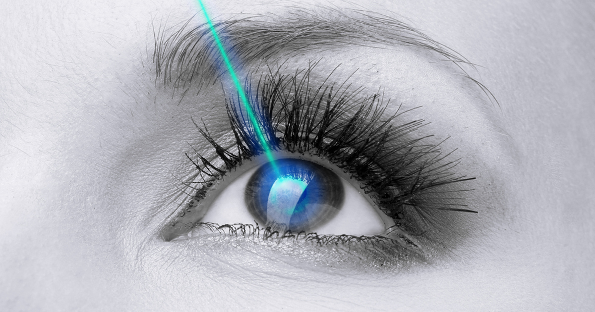
Laser-assisted in situ keratomileusis, better known as LASIK is a type of laser refractive surgery. It is by far the best known and most commonly performed eye corrective surgery. In general, a special type of cutting laser is used to precisely change the shape of your cornea to improve vision. In a normal functioning eye, images are clearly focused on the retina because the light rays are bent properly to contact the retinal surface. With nearsightedness, farsightedness, or astigmatism, the light is bent incorrectly and it ends up being focused in the wrong location, resulting in blurred vision. A person can use contact lenses or glasses to refract the light correctly, but reshaping the cornea itself also will provide the necessary refraction.
Related Article: Vision Care
Lasers have not been in existence for a long time. Imagine telling the person that invented lasers that we would be using them on an eyeball in the future. Like any surgical procedure, there are risks. Consult with a doctor about LASIK if you feel it will help any issues you are having.
Wave Scanning
Wave scanning is a is a pre-test for LASIK. Many people suffer with nearsightedness, farsightedness, or astigmatism. Even if those are corrected, the human eye will still have some subtle distortions in their vision. Wave scanning technology measures the subtle distortions and adjusts the LASIK treatment to account for them. Studies show, with wave scanning-guided treatments, a higher percentage of subjects achieved 20/20 vision than with LASIK alone. LASIK is a fantastic procedure, but coupling it with wave scanning technology makes it even more effective.
PRK
PRK or Photorefractive keratectomy was the first laser eye surgery developed that reshapes the cornea. Instead of cutting the cornea, like LASIK, the PRK procedure involves removing the outer layer of the cornea prior to reshaping it with a laser. PRK requires a longer recovery time than LASIK, but it is better for some patients. Results are about the same with PRK and LASIK. Deciding what surgery will work for any given patient often depends on which pre-existing conditions are present. PRK does not make any changes to human eye anatomy because the eye can grow back those cells it lost in a few days.
RLE
Refractive Lens Exchange (RLE) is a surgery that will actually change the anatomy of the human eye. With RLE, the lens that an individual was born with will be replaced. The lens is replaced with an artificial intraocular lens. The new lens corrects refractive issues and is designed to help patients with farsightedness.
KAMRA Inlay
The KAMRA Inlay eliminates the need for reading glasses for patients that lose their ability to read up close because of age. In a brief, 10-minute, surgical procedure, a doctor uses a laser to create a small pocket or flap in the cornea. A tiny inlay is inserted under a flap in the center of the cornea. The inlay is only about 0.15 inch in diameter and has a small pinhole in the middle. This allows focused light into the eye. The inlay is usually inserted into the patient’s non-dominant eye, and the brain allows both eyes to work together to give the patient clearer vision. Although it rarely happens, if the inlay causes problems, it can easily be removed.
GVRSS
Gentle Vision Retainer Shaping System is a non-surgical option. GVRSS uses specially designed vision retainer lenses similar to contact lenses to gently and gradually reshape the cornea. It is worn while sleeping to aid in providing patients with clear vision by eliminating or reducing nearsightedness or astigmatism.
Blindness
Currently, there is no cure for blindness. However, with advances in technology, several institutions are getting close to restoring vision. Some of these innovations suggest an eye transplant. A transplant wouldn’t change human eye anatomy to correct blindness, but it would require changing the person. Other studies suggest using stem cells to cure blindness. There are no certain cures for blindness, but technology is certainly making it easier to search for a cure.
Conclusion
Technology with its blue light and LEDs is not going away in the future. Neither is our dependence upon it. The best thing an individual can do to avoid any type of negative consequences from technology is to take regular breaks. We are only given one set of eyes and technology has not gotten humankind to the point where eyes can be replaced. If the proper precautions are taken and only slight corrections need to be made, modern technology can have a positive effect on our eyes. Regular check-ups can help avoid any major issues that may require invasive surgery or the loss of eyesight completely.
