Although the eyes do not make up much of a human’s overall mass, they are extremely important when it comes to the tasks we do on a daily basis. Since the beginning of time, eyes have been a source of fascination. Colored lenses have been discovered in ancient Greece and the region now known as the Netherlands. No conclusive data has shown when eyeglasses were invented, but some believe the ancient Chinese developed corrective lenses. Knowing that attempting to correct vision occurred so long ago shows the importance of our eyes. Although adaptive devices have come a long way in recent history, nothing has been able to reproduce sight for those that were born blind or lost their vision. This article will explore eye parts and functions. It will also explore some factors that affect the human eye and its functions.
There are twelve major parts of the eyes. Looking into a person’s eyes, it may seem that the eye is simple. The lids open and close, the pupil dilates, and the eye moves to see our environment. On a very elementary level, that describes the eye perfectly. However, this article will explore how the many parts of the eye work together and allow humans to see. We will explore each part’s function and how it is constructed.
Top Related Article: Symptoms and Signs of Eye Health
So, what is the function of the eye? In this article, imagine that the eye has been cut in half vertically. The eye will be described from the front, starting at the cornea, to the back, finishing at the optic nerve. At the end of this article, after the eye has been described, a few ailments and other things that affect the eye will be covered. Let us start with the first thing a person sees when they look another person in the eyes. Although a person may never actually see the cornea, it is the first thing they will look through.
Eye Function: Eye Parts and Functions
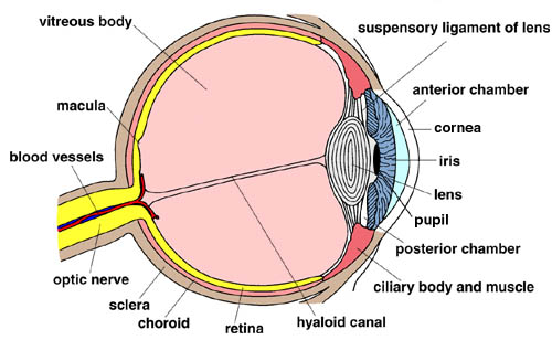
The cornea is the clear, front surface of the eye. The cornea appears wider than it is tall. This is caused by the sclera covering some of the top and bottom of the cornea. The cornea consists of the five layers. The first layer is the corneal epithelium. The cells of the corneal epithelium are constantly being stripped away and replaced. The second layer is known as Bowman’s layer. The Bowman’s layer consists of dense, fibrous connective tissues. This layer forms the transition between the corneal epithelium and the stroma. The next layer is the corneal stroma. The corneal stroma represents about 90% of the cornea’s thickness. This layer consists of bundles of collagen fibrils, known as lamellae, that are uniformly spaced. The uniform spacing of these lamellae allows the cornea to be perfectly clear. The fourth layer of the cornea is Descemet’s membrane. This membrane separates the stroma from the endothelial layer. The inner most layer of the cornea is known as the corneal endothelium. This layer is covered in aqueous humor. This aqueous humor fills the space between the cornea, the iris, and the pupil.
Related: Eye Anatomy
The function of the cornea is rather simple. The cornea’s main function is to focus the eye. The cornea provides 65-75% of the eye’s focusing power. The cornea is able to do this by allowing light to enter the pupil. The next part of the eye we will cover is often used to describe other people.
Iris
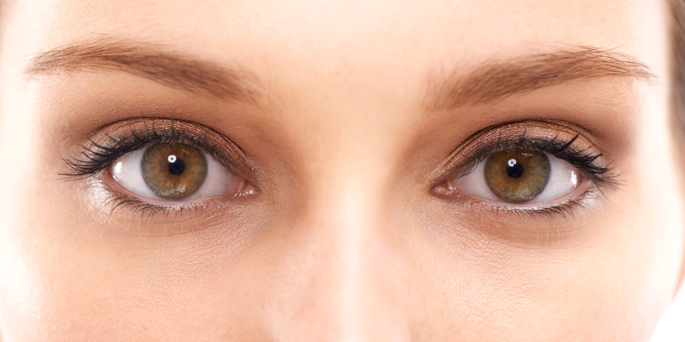
The iris is a thin, circular structure directly behind the cornea. The iris is the most visible part of the eye. This small structure is responsible for a person’s eye color. Everyone has the same color of melanin pigment in their iris. Color is determined by the amount of melanin pigment in a person’s iris. A person with blue eyes has much less pigment than a person with brown eyes. The iris is heavily pigmented so that light cannot shine through it.
The iris consists of two layers. The first layer, known as the stroma, consists of pigmented fibrovascular cells. The stroma connects to the sphincter that controls the pupil size. They next layer is made of pigmented epithelial cells. This layer is where most of the pigment is located so that the only place light can enter the eye is the pupil. The iris is also divided in to two regions. The pupillary zone forms the edge of the pupil. The ciliary zone consists of the remaining parts of the iris. The ciliary zone extends from the pupillary zone to the ciliary body of the eye. There are two major structures that assist the iris. The Crypts of Fuchs allow the stroma to be coated in aqueous humor. The radial contraction folds of Schwalbe are associated with the scalloped appearance of the pupillary ruff.
Much like the cornea, the function of the iris is simple. The function of the iris may be simple, but it is extremely important. The iris’ sole function is to control the diameter and size of the pupil. Although only two parts of the eye have been listed, aqueous humor has been mentioned as something that both parts use. The next part of the eye is responsible for that aqueous humor.
Ciliary Body
The ciliary body is a circular structure that is an extension of the iris. This structure secretes aqueous humor via the ciliary processes. It is also part of the uvea which is a vascular system in the eye that provides oxygen and nutrients to the eye. The ciliary body contains the ciliary muscle that is responsible for changing the shape of the lens. Changing the shape of the lens allows the eye to focus on a near object. This process is known as accommodation. The next part of the eye isn’t even a part of the eye. It is actually a void where no parts of the eye touch.
Pupil
Technically speaking, the pupil is part of the eye. However, the pupil isn’t even a structure in the eye even though it is very important to eye function. It is simply the open space in the center of the iris. The pupil allows light to enter the eye so that the light can be focused on the retina. Once the light focuses on the retina, the process of sight has officially started. The pupil appears black as the light that travels through the pupil is absorbed by the retina and not reflected. If the pupil appears cloudy or pale, that discoloration is caused by a cataract on the lens. A common misconception about the pupil exists when taking flash photography. Sometimes a picture shows that an individual’s pupils are red. This is caused by too much light hitting the pigment in the choroid. The pigment that protects the choroid from absorbing light and limiting reflection actually causes that red eye reflection.
Related Article: Vision Care
The size of the pupil is controlled by the muscles of the iris. When the pupil dilates in low light situations, the process is known as mydriasis. When the pupil constricts in bright light, the process is known as miosis. An interesting fact about the pupil is that the size varies from person to person. As noted, the pupil is not a structure at all. The next part of the eye to be discussed is the biggest structure of the eye.
Sclera
The sclera is the largest part of the eye and is extremely important to eye function. It is the white part of the eye that surrounds the cornea. The sclera is 80% of the eye’s surface. Dense connective tissue of the sclera is connected to the stroma layer of the cornea. This tissue allows for the strength and flexibility of the eye. When looking at the white of someone’s eye, you could see blood vessels. One might assume that those blood vessels belong to the sclera; however, the sclera lacks blood vessels entirely. The sclera receives its nourishment from the episclera or choroid.
The sclera has a few very important functions. It is responsible for maintaining the shape of the eyeball. It also protects the eye against external trauma. Another feature of the sclera is that it is the attachment point for the extraocular muscles that control the movement of the eye. The next part of the eye helps protect the sclera and cornea.
Conjunctiva
The conjunctiva is a thin, clear membrane that covers the front surface of the eye and the inner surface of the eyelids. The bulbar conjunctiva covers the anterior surface of the sclera, but does not cover the cornea. The palpebral conjunctiva covers the inner surface of the upper and lower eyelids. The bulbar and palpebral conjunctiva are one continuous segment. Being one continuous segment prevents contact lenses from being lost behind the eye.
The conjunctiva serves the eye in several different ways. It keeps the eye moist and lubricated. Along with lubricating the eye, it lubricates the inner eyelids to prevent friction. The conjunctiva protects the eye from dust, debris, and infection-causing microorganisms. It has special cells that secrete a component of tears to prevent dry eye syndrome. Another task it completes is that it delivers nutrients to the eye and the eyelids. The next part of the eye isn’t about protecting structures within the eye, it is all about sight.
Lens
The lens is a transparent structure that is located directly behind the iris. Unlike the lens of a camera, this lens can change shape to focus on different objects. By changing its shape, the lens is able to bend light rays so that they form a clear image on the retina, which is central to eye function. The ciliary muscles attach to the lens to change it shape and curvature. The lens provides 25-35% of the eye’s focusing power, but weakens with age. As the lens ages, it loses elasticity and is unable to change enough to focus on objects. This is why older individuals often use reading glasses.
The lens is divided in to three parts. The lens capsule is very elastic, it allows the lens to assume its globular shape. The next part is the lens epithelium. The lens epithelium produces new fibers so the lens continues growing throughout its life. The next part is the lens fibers. These fibers make up the bulk of the lens. The next part of the eye is where the lens focuses any light that it receives.
Macula
The macula is a small, but highly functional part of the retina. It is directly in the center of the retina. The macula is the point where all light is focused. Once the light is there, the macula will take a “picture” and send that picture to the brain where vision is completed. The macula is very important as it provides sharp, central vision that we need for reading, driving, and seeing fine detail. The remaining part of the retina provides humans with peripheral vision. Located on the macula is an even more important part of the eye.
Fovea Centralis
The fovea centralis is a depression in the center of the macula. When light is focused on this depression, eyesight is the sharpest. Although the fovea centralis is only 1% of the retina’s overall size, it takes 50% of the visual cortex of the brain. It is the only area in the retina where 20/20 vision is attainable. The fovea centralis is extremely small, but plays a huge role in eyesight.
Retina
The retina is located at the back of the eye. It is the sensory membrane that lines the inner surface of the back of the eyeball. It is full of photoreceptor cells known as rods and cones. Rods are responsible for vision at low light or scotopic vision. They are not responsible for color vision and have a low spatial acuity. Cones are active at high light levels or photopic vision. They are responsible or color vision and have a high spatial acuity. The fovea centralis is completely populated by cones. Mesopic vision is the light level in which both rods and cones are fully operational.
The photoreceptor cells of the retina take light focused by the cornea and the lens and convert it into chemical and nervous signals. These signals are sent back to the brain. The next part of the eye provides precious nutrients to the retina. The proper blood flow and nutrient balance are absolutely necessary for the retina to function properly.
Choroid
The choroid is the vascular layer of the eye. The choroid has been mentioned several times throughout this article because it does play a vital function in the eye. It provides all of the blood flow to the retina. The choroid is located between the retina and the sclera. The choroid consists of the four layers. The Haller’s layer contains the large blood vessels while the Sattler’s layer contains medium sized blood vessels. The choriocapillaris contains the capillaries and the Bruch’s membrane is the innermost part of the choroid.
The choroid is important when developing vision as a child as it can move the retina. The choroid also secretes substances that are thought to be involved in the growth of the sclera. Much like the pupil, the next part of the eye really isn’t part of the eye at all.
Optic Nerve
The optic nerve is attached to the rear of the eyeball. This nerve connects the eyeball to the brain. It is responsible for carrying impulses from the retina to the brain. When these impulses arrive at the brain it interprets them as images. The optic nerve consists of over 1,000,000 nerve fibers. Just as the pupil is not a structure in the eye, the optic nerve is considered a part of the central nervous system and not a part of the eye.
Every part of the human eye plays an integral role in eye sight. The eye itself is protected by eyelids, eyelashes, and has some built in protection too. Unfortunately, there are many things that can affect the eye. No part of the eye is 100% safe from being affected. The next section of this article will focus on diseases or disfunction within the eye that can cause major issues.
Factors That Affect the Eyes
Corneal Abrasion
One of the most common eye injuries is the corneal abrasion. A corneal abrasion is a scratch or a cut on the outermost surface of the cornea. These abrasions can be caused by an incidental finger poke or by dirt and sand. Generally, a corneal abrasion does not require a visit to the doctor. This type of injury can be soothed with clean water or saline solution. There are many holistic approaches to cleaning your eye.
Related: Treatment Options for Eye Conditions
Corneal Ulcer
A corneal ulcer is an open sore on the cornea. The damage occurs in the epithelial layer. A corneal ulcer usually results from an eye infection, but severe dry eye can be a cause as well. A corneal ulcer is due to the invasion of bacteria, fungi, or viruses. Often a corneal ulcer can be treated with topical antimicrobials and dilating drops. An ophthalmologist visit is required to get the proper medications to treat the ulcer.
Cataracts
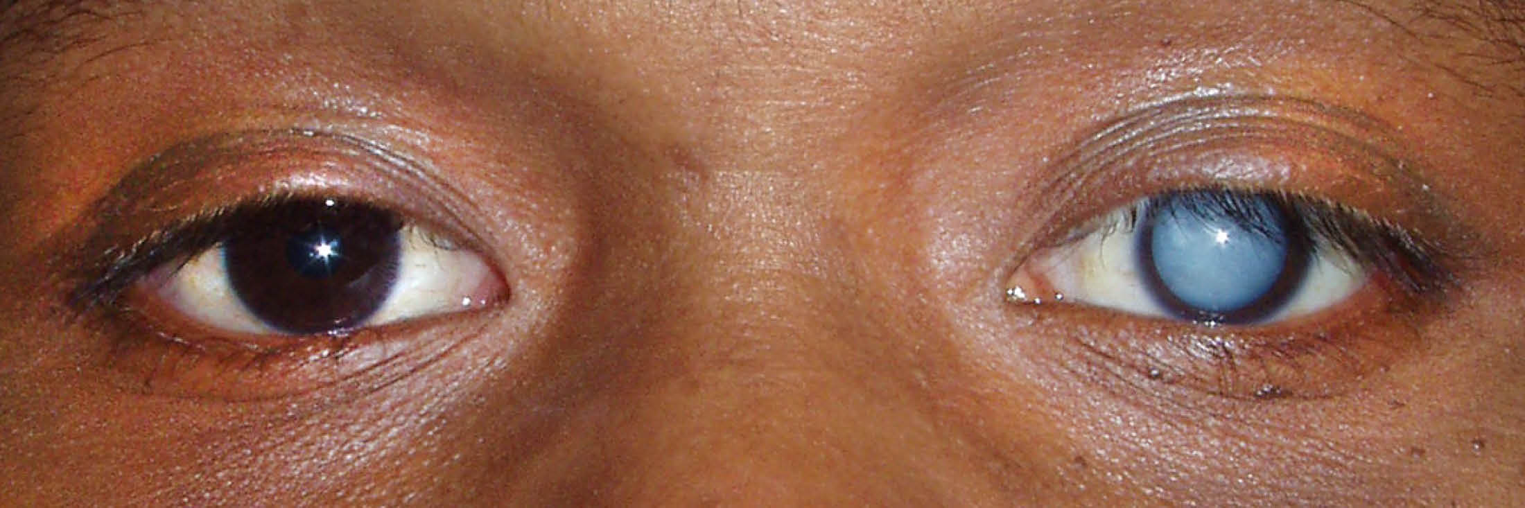
A cataract is a clouding of the normally clear lens of your eye. For people who have cataracts, seeing through cloudy lenses is a like looking through a foggy window. Clouded vision caused by cataracts can make it more difficult to read, drive a car, or see the expression on a friend’s face. Cataracts form on the lens of the eye. As noted earlier in this article, the lens becomes less flexible, less transparent, and thicker with age. Tissue within the lens begins to break down and clump together which causes clouds on the lens. Generally, if a person has a cataract in one eye, they will have a cataract in the other. They will not develop at the same speed so there will be a difference in vision between the eyes.
Cataracts can develop for multiple reasons. Most cataracts develop slowly and don’t disturb your eyesight early on. But with time, cataracts will eventually interfere with your vision. Some cataracts develop after an injury to the tissue that makes up the lens of the eye. If cataracts are left untreated, they will become bigger and denser.
There are four types of cataracts. Three types are named for their location, nuclear, posterior, and cortical. Congenital cataracts are cataracts that you are born with or develop during childhood. At first, stronger lighting and eyeglasses can help you deal with cataracts. But if impaired vision interferes with your usual activities, you might need cataract surgery. Fortunately, cataract surgery is generally a safe, effective procedure.
If a person is concerned about developing cataracts, they can quit smoking, wear sunglasses more often and reduce their consumption of alcohol.
Glaucoma
Glaucoma is a disease that damages your eye’s optic nerve. It usually happens when fluid builds up in the front part of your eye. The extra fluid causes an increase of pressure in your eye. This pressure, if left untreated, will damage the optic nerve. Glaucoma is the leading cause of blindness in people over the age of 60. Glaucoma is the second-leading cause of blindness in the United States and second-leading in the world. With early detection and treatment, glaucoma can be prevented.
Unfortunately, early detection is difficult. Early stage glaucoma has no symptoms. By the time glaucoma is discovered, irreversible vision loss has occurred. One cause of glaucoma is ocular hypertension. Ocular hypertension results when you have higher-than-normal pressure inside the eye. Glaucoma usually interferes with peripheral vision before causing complete blindness.
There are two major types of glaucoma. Each type of glaucoma refers to the angle in which aqueous humor drains from the eye. If the aqueous can access the drainage angle, the glaucoma is known as open angle glaucoma. If the drainage angle is blocked and the aqueous cannot reach it, the glaucoma is known as narrow angle glaucoma.
There are several procedures that can help treat glaucoma. Depending on the severity of a person’s glaucoma, they can have glaucoma surgery. If glaucoma is not too severe, lasers and medication can be used as a treatment. If detected early, eye drops can be used to reduce the eye pressure. There is no clear evidence that anything can be done to prevent glaucoma. Some studies have concluded that exercise can reduce a person’s risk of glaucoma. As always, routine eye exams can detect any type of potential glaucoma causing issues.
Color Blindness
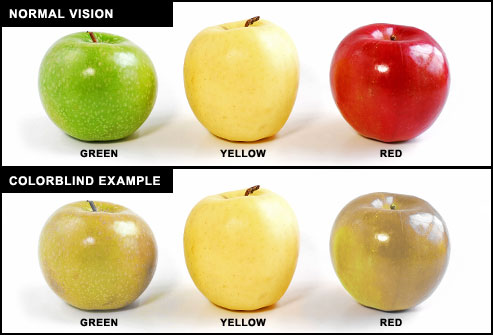
Color blindness is a bit of a misnomer as it is not a form of blindness at all. It is simply a deficiency in the way a person sees color. Interestingly, color blindness is an inherited condition that affects males more frequently than females. Color blindness occurs when light-sensitive cells in the retina fail to sense variations in light wavelengths. As noted earlier, rods and cones make up the photoreceptor cells in the retina. Rods cannot sense color, so color blindness is caused by deficiencies or the absence of cones in the retina.
Currently, there are no ways to prevent color blindness in humans. Gene therapy has been used to cure color blindness in monkeys, but nothing has made the cure possible in humans. The best way for a person to deal with color blindness is to simply adapt their life style. They may not qualify for jobs that require discerning colors for safety reasons. An eye exam can determine if special lenses could be ordered to help the individual overcome their color blindness.
Macular Degeneration
Macular Degeneration is the leading cause of vision loss, affecting more than 10 million Americans. It affects more Americans than cataracts and glaucoma combined. Macular Degeneration is an incurable eye disease and is caused by the deterioration of the central portion of the retina or the macula. When it is working properly, the macula collects highly detailed images at the center of the field of vision and sends them up the optic nerve to the brain, which interprets them as sight. As the cells of the macula deteriorate, images are not received correctly. Macular Degeneration begins with wavy or blurred vision. As it progresses, central vision is completely lost. Although a person with macular degeneration maintains their peripheral vision, they are considered legally blind.
There are three stages of age-related macular degeneration (AMD). Early AMD does not have any amount of vision loss. AMD can be detected at this stage by finding yellow deposits beneath the retina. Intermediate AMD does have some vision loss, but it may not be noticeable. Comprehensive eye exams can detect AMD with more tests of the retina. Late AMD is marked by noticeable vision loss. There is still no conclusive evidence of what causes the cells of the macula to degenerate. As with all eye related damage and diseases. Routine eye exams can help with early detection and treatment.
Retinal Detachment
Retinal detachment describes an emergency situation in which the retina pulls away from its normal position. It’s the separation of the retinal cells from the layer of blood vessels that provides oxygen and nourishment. The longer retinal detachment goes untreated, the greater your risk of permanent vision loss in the affected eye. Retinal detachment is painless, but there are ways to detect it. Flashes of light in one or both eyes, the appearance of floaters, blurred vision, and a reduction in peripheral vision are all signs of a detached retina. If a person suspects they have a detached retina, immediate medical attention is necessary.
There are three different types of retinal detachment. Rhegmatogenous is the most common. This occurs when a small hole opens in the retina and fluid is builds behind it. The buildup of fluid causes the retina to pull away from the underlying tissues. The second type of retinal detachment is tractional. If scar tissue grows on the retina’s surface, it can cause the retina to pull away. This is often caused by uncontrolled diabetes. The last type of retinal detachment is exudative. Much like rhegmatogenous, exudative retinal detachment is caused by a build up of fluid behind the retina. There is no hole or tear, so macular degeneration or an eye injury can lead to exudative retinal detachment. Immediate medical attention can prevent permanent vision loss due to a retinal detachment.
Conclusion
The human eye is a complex system that seems so simple. Most of us open our eyes in the morning and aren’t even aware of how the eye works. Our eyes are extremely important and should be well take care of. There are a multitude of diseases and accidents that can temporarily or permanently affect our vision. Wearing the proper safety equipment can protect our eyes at work and routine eye exams can detect any diseases that we may contract. It is important to understand how our eyes work in order to protect them the best we can.
