The more you learn about the structure of your eye, the easier it will be to properly care for these important organs and your entire visual system. Eye structure is a fascinating area of medical inquiry which has expanded over the years in many ways. In this article, we’ll go in depth exploring the structure of the human eye so that you can be better informed and know how to properly approach any eye issues you may be facing.
To better understand the structure of your eye, the best place to start is understanding the parts of the eye. Below is a list of the major parts of the eyeball, which is about a one inch thickness. Then, the eyeballs are connected to the optic nerve, which is connected to many other parts of the brain that make up the entire visual system. We’ll start with the anatomy and the structure of eyeball, and then continue to explore the other parts of the entire visual system as well.
Top Related Article: Symptoms and Signs of Eye Health
The eyes are known as paired structures in anatomical terms. This means that they offer slightly different interpretations of the image in front of your eyes, and then your visual cortex must interpret those images to make sense of what’s right in front of your face. Another way our visual system is described is that humans have binocular vision. This means that humans can perceive 3D images that make it possible to have depth perception.
How the Eye Works
If you have an understanding of how a camera works, then you’ll have a good idea how your eye works. Of course, when photographers were beginning to develop the camera, they modeled it after their understanding of how the eye functioned.
 Light is what makes it possible for the eye to see things in the visual field. The cornea is the clear surface of the eye that acts like a camera lens. Light is focused by the cornea, making sure it enters the eye appropriately.
Then, the iris of the eye controls the amount of light reaching to the back of the eye. It does this by automatically adjusting the size of the pupil. The pupil can be understood like the aperture of a camera.
The eye’s crystalline lens is found right behind the pupil. It helps to focus the light even further after the cornea initially focuses it. The focusing that the eye’s lens does is called accommodation. It can help the eye focus on objects that are very near or getting closer. It works kind of like the autofocus on a camera lens.
The light that is focused by the two lenses, the cornea and the crystalline lens, then makes its way to the retina. The retina is a very sensitive part of the eye and plays a very unique role in allowing you to see. Basically, the retina is a lining of cells that are photosensitive and can tell whether light is being sent to the eye or not.
Top Related Article: Human Eye Anatomy And How Eyes Are Impacted By Technology
Basically, it works like an image sensor. If you had a digital camera, your camera would have an electronic image sensor that could sense when there was an image in view. The retina works in a similar way. Once it senses the image by interpreting the light that enters the eye, the retinal cells then translate that image into electronic signals. From there, the signals are transmitted to the visual cortex.
Three Chambers of the Eye
One way of thinking about the way the structure of eye works is by understanding that there are three chambers of the eye. These chambers could be considered sections of the eye. When you understand what the three chambers are made of, you can have a more firm understanding of the eye in general.
The three chambers of the eye include the anterior chamber, the posterior chamber, and the vitreous chamber. The anterior chamber of the eye is the area between the cornea and the iris. This is the smallest chamber of the eye. In this chamber, light is focused by the cornea and the contraction of the iris around the pupil, where light enters the eye.
The posterior chamber is the area between the iris and the lens. This is a slightly larger chamber where light is refracted through the lens and refocused. And the vitreous chamber is the area between the lens and the retina. In this chamber of the eye, the light passes through the fluid in the vitreous area and is sent to the retina, where special photoreceptor cells pick up on the images being processed through the eye.
The Basic Structure of the Eye
Here are the structures of the eye:
>Iris
>Cornea
>Pupil
>Lens
>Vitreous Body
>Optic Nerve
>Ciliary Body & Muscle
>Anterior Chamber
>Sclera
>Choroid
>Retina
>Macula
Once you have an understanding of all these parts of the eye, you can have a sense of the entire structure of the eye as well.
Top Related Article: Eye Function And Factors That Effect Eyes
The Pupil
Believe it or not, the most important part of the eye doesn’t have any cells associated with it. In some sense, it’s not really a structure at all–it’s more like an open space. The pupil is found in the center of the iris. The iris is basically the structure of cells that is found around the eye.
The pupil plays a vital role in vision: it is where light enters the eye. Without an opening in your eyes, there would be no way for you to see. Before light enters the eye, it passes through the cornea, which influences the way that it is processed. But once the light enters the eye through the pupil, it is processed and sent to the retina.
Pupils are perfectly round and typically have an equal shape to one another. The pupil appears black because the light that hits the pupil is not reflected back through the retina.
Have you ever wondered why people sometime show up on camera having very bright color eyes–that are abnormal? The “red-eye” effect in camera is caused by the pupil! When a camera takes a picture of the eye, the flash reflects back through the retina, and the bright red color is shown.
The pupil is a commonly examined part of the eye that can tell us a lot about someone’s general visual health. Eye doctors will test how easily someone’s pupil expands and contracts in order to determine the overall health of their visual system.
The Uvea
The uvea is known as the middle layer of the eye. It is the pigmented layer of the eye as well. It consists of an iris, a ciliary body, and the choroid. These three parts have specific functions that help the entire visual system and form an essential aspect of the structure of the eye.
Iris
 In some ways, the iris is the most recognizable part of the eye. It is the part of the eyeball that gives your eye its color. The iris is the ring of tissue that appears behind the cornea. The iris expands and contracts to adjust the size of the pupil. This regulates how much light enters the eye. In low light situations, the iris contracts, making the pupil larger. In very bright situations, the iris expands, making the pupil smaller.
The iris is a circular structure that is quite thin. It is made up of connective tissue and muscle that surrounds the pupil.
There are two known layers of the iris. One layer keeps light from reaching the retina when it is not necessary for understanding the image in the visual field. The other layer is the layer that contains the cells that make eye color possible. These cells are called chromatophores. The concentration of melanin in the chromatophores is what gives the iris its various colors. If you have a high concentration of melanin in your chromatophores, you’ll have darker colored eyes. If you have a low concentration of melanin in your chromatophores, you’ll have light colored eyes.
Ciliary Body
The ciliary body is known as the second part of the uvea. It is located behind the opaque sclera of the eye. It can’t be seen because it is being blocked by the sclera of the eyeball. Additionally, it surrounds the iris. The ciliary body has an important role in the structure of the eyeball. It’s connected to a network of ligaments called the zonules of Zinn. Another name for these ligaments is called the ciliary zonules. This part of the eye secretes fluid that fills the space between the iris and the lens and the cornea of the eye.
The ciliary muscle is part of the ciliary body. This muscle is what makes it possible for the lens to accommodate the changes in the visual field, shifting its shape so that it is capable of sending light into the retina. The ciliary body is also responsible for secreting a fluid called the aqueous humour that is supportive to the lens and the iris.
Choroid
The choroid is the third part of the uvea. It contains many very small blood vessels surrounding the retina. These blood vessels serve to nourish the retina, and are essential for maintaining the health of your retina. That’s a vital part of keeping your entire visual system healthy, because the retina is essential to having good eyesight.
Sclera
The sclera is the white part of your eyes that surrounds your cornea. When you look the eyes of someone you know, the whites of the eye only take up a small percentage of the eyeball. But the reality is that the sclera takes up more than eighty percent of the eyeball. Collagen and fibrils are arranged in many bundles. This is what makes the eyeball so strong and flexible.
The sclera has a very limited blood supply. It can range in thickness from one millimeter to three-tenths millimeters. The sclera is nourished by blood vessels in the episclera, a loose layer of tissues that is found underneath the conjunctiva.
The central function of the sclera is to maintain the shape of the eyeball, essentially playing a structural role in the eyeball. It’s a tough, fibrous part of the eyeball and can protect more sensitive parts of the eyeball from external trauma. It’s also the place where all of the ocular muscles can attach and regulate the movement of the eye.
Vitreous Body
The vitreous body takes up more space in the eyeball than any other part of the structure of the eyeball. The vitreous body is basically what fills the body of the eyeball. It’s a clear, jelly-like substance that fills the entire eye. It can be found from the lens of the eye to the back of the eye.
The vitreous body plays other important roles in keeping the visual system functioning at a high level. For one, the vitreous body contains a fluid called the vitreous humour. This fluid allows the eyeball to absorb shock, especially to the retina. This shock can arise during rapid eye movements, especially during sleep. This part of the eye also helps to ensure that the lens stays in the appropriate position. The shape and pressure of the vitreous body also ensures that the retina is smooth against the rear of the eyeball. This is a very important job, because if the retina is not properly placed against the wall of the eyeball, you could experience retinal detachment, which can seriously impair your vision.
Cornea
 The cornea is the front of the eyeball. It plays an essential role in the structure of the eye. It’s found in front of the iris and pupil and it is what allows light to enter the eye. From the front of the eye, the cornea appears to have more width than breadth. Typically the cornea is about twelve millimeters wide and eleven millimeters high. In relative terms, it is about two-thirds the size of a dime.
The cornea plays a very important role in the visual system. That’s because it is responsible for allowing light into the eye for vision. It also contributes to the ability of the eye to focus on objects. The issues that many people have with their vision, including refractive errors, astigmatism, farsightedness, and nearsightedness, all have to do with the cornea.
There are five layers to the cornea, and they include:
>The Corneal Epithelium
>Bowman’s layer
>The Corneal Stroma
>Descemet’s membrane
>The Corneal Endothelium
The Corneal Epithelium
This is the outer layer of the cornea. It is about five or seven cells thick. This means it is about ten percent of the cornea. This epithelium is made up of epithelial cells that are constantly produced and then shed from the surface of the eye. This means that the corneal epithelium is completely replaced within in one week. The role of this layer of the cornea is to provide the right surface for tears to spread across the eye, keeping the eye healthy and vision clear.
Bowman’s Layer
This is a very thin layer of the cornea. It is basically a transitional layer between the corneal epithelium and the stroma that is located underneath. It is a dense layer that helps to protect the cornea from scratches, especially the more sensitive layers that are found underneath this layer. In general, as long as any corneal scratches don’t reach the Bowman’s layer and only penetrate the corneal epithelial layer, you can usually recover fully.
The Corneal Endothelium
This is the deepest layer of the cornea. The endothelium is a layer of cells that only measure approximately five microns. The cells in this layer are irregularly shaped and are arranged in interesting patterns that can sometimes be called an endothelial mosaic. This is where the fluid content of the cornea is contained. If you damage this part of your cornea, you can end up with an edema and experience swelling.
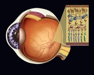
Retina
The retina plays an essential role in the visual process. The retina is a sensory membrane that lines the inner surface of the eyeball. It has many layers of cells that make it possible for you to create images, and is composed of special cells that are called photoreceptors. These special cells are specially designed to respond to light exposure.
There are two types of photoreceptor cells that are called rods and cones. The rods are the cells that can detect motion. They allow for you to see things in black and white, and they assist you in function in low light. The cones are the cells that make it possible for you to see in color and they also offer central vision.
Rods are cells that can be found throughout the retina. Cones are centralized in a small area called the macula. At the very center of those cones is a small depressed area called the fovea. The fovea is made up only cones and is the place in the eye that has the highest level of visual acuity.
The photoreceptor cells in the retina are able to receive the light that is let in by the cornea and the lens. Then they can translate that light into nervous and chemical signals. Those signals are then taken up by the optic nerve and transported into the visual centers of the brain.
Other Structures of Human Eye
Alongside the structures and parts that make up the eyeball itself, there are many other parts to the eye. These structures help protect and clean the eyeball so that all of the other structures of the eye can keep functioning at a very high level. These extra structures include:
>Eyelid
>Lacrimal Gland
>Eyelashes
>Lacrimal puncta
>Lacrimal Duct
>Inferior Lacrimal Canal
>Conjunctiva
Top Related Article: Eye Anatomy | Everything You Should Know About Your Eyes
Eyelid
The eyelid helps primarily to protect the eye. It’s a loose layer of skin that allows for the eyelids to move freely. There are also a couple types of glands around the eyelids that secrete a substance that interacts with the tears to keep the eye properly lubricated as much as possible.
Eyelashes
The eyelashes keep debris and dirt out of the eye. They are kind of like the windshield wipers on your car. When small bits of dirt or debris want to enter the eye, the eyelashes keep them at bay.
Lacrimal Gland
This is the gland in the body that produces the tears around your eyes. There are actually a lot of different ingredients in your tears. The ingredients of your tears include proteins, metabolites, electrolytes, lipids, and enzymes.
Conjunctiva
The conjunctiva is the inner lining around the eye socket. It has special cells called goblet cells that help to create the recipe of your tears.
Learning about the structure of your eye can help you understand how your visual cortex works. The more you take in about how the visual cortex works and what it can do to help your vision in general, the better off you’ll be in terms of maintaining the structure of eye, and ensuring that your vision works well throughout your life.
Once you have a firm understanding of the structure of eye, you can then learn more about how these structures are connected to all of the important organs in the face and head. The more in-depth you go with your understanding, the better off you will be as you consider how to move forward with your overall eye health. So if you’ve ever wondered, what is the structure of the eye, look no further than this article so you can fully understand it and incorporate it into your overall knowledge about your body’s structures!
