
[vc_row][vc_column][vc_column_text]By be tter understanding about the eye and how the eyes work, you can identify when something isn’t right. The five senses: sight, hearing, taste, smell, and touch, allow us to better understand our surroundings, the world, and even ourselves. We are able to gather and process a wealth of data and information through the five senses. Of these five senses, many people argue that vision is the most important, and many people also report that they would fear to lose vision the most.
Now, why might people feel this way about vision? Just take a brief moment to imagine trying to live your everyday life without being able to see. You couldn’t see the sunlight through your curtains telling you it’s morning. How would you be able to walk out of your room and begin getting ready for your day? Clearly, it’d be a difficult thing, and your perception of the world would be drastically different if you couldn’t see. In fact, it is reported that more than 75% of the information we receive and process about the world is through sight. Therefore, it’s important to know about the eye and how the eyes work.
Top Related Article: Human Eye Anatomy And How Eyes Are Impacted By Technology
Visualization of the world occurs through our eyes, an organ that is often overlooked in its importance and complexity. For simplicity’s sake and to better understand how this crucial organ works, it is often described analogous to the human version of a camera. Each eye operates individually gathering the sensation of light and other visual cues and processing this information to provide our perception of vision. In this article, we will cover the important anatomy of the eye and further focus on how the human eye works, as well as the individual functions that allow us to better see the world. It is important to note that the anatomy of the eye is more in-depth than what is covered in this article, but if you would like to dive deeper into that content, Eye Anatomy also written on this site.
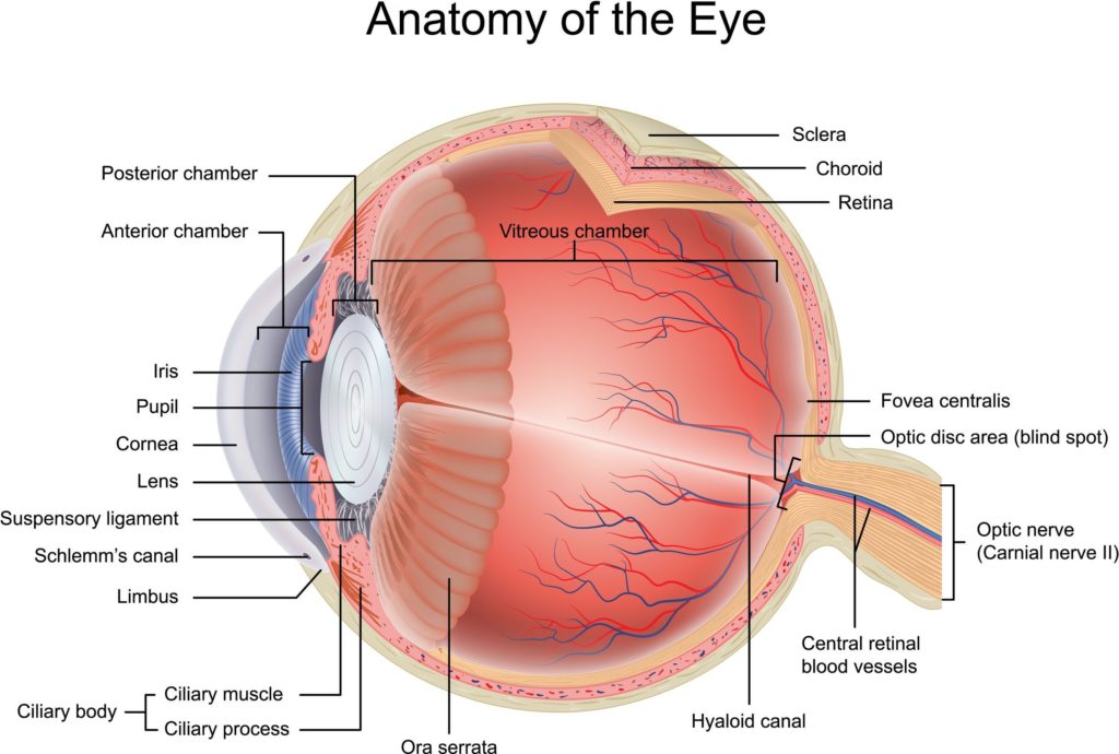
Before we dive into how the eye works and the associated parts that are involved in sight, we’ll briefly cover the anatomy that surrounds the eye. There are numerous anatomical structures, which include the orbit, eyelids, eyelashes, and extraocular muscles. The orbit is the bony socket in which each eye lies in your skull. It is comprised of multiple bones, including the cheekbone, forehead, temple, and side of the nose. The orbit also houses the multiple muscles involved in moving the eyes, the blood vessels and nerves involved in the eye. The eyelids serve as an additional protective layer for the eye, preventing foreign matter, which includes dust, dirt, foreign objects, from entering your eye and leading to damage. The eyelids also act to spread tears and lubrication over the surface of your eye. Similarly, your eyelashes also act as another protective layer.
We will now briefly mention that there are extraocular muscles that surround the eye and allow it to move and rotate across different planes. All you need to know for the purposes of how vision works is that there are six of these extraocular muscles that allow the eye to move freely.
How the Eyes Work: Different Parts of the Eye
Now we will begin our discussion on the anatomy of the eye itself and discuss the structures that are pertinent to our perception of vision.
Conjunctiva
This is a transparent layer that covers the surface of the eye and the inner surface of the eyelids. It acts as an additional protective layer. There are two segments of the conjunctiva: the bulbar and palpebral conjunctiva. The bulbar conjunctiva covers the anterior part of the sclera. It does not cover the cornea; it extends to the point where the sclera and cornea meet. The palpebral conjunctiva covers the inner portions of the eyelid on each eye. Because the two palpebral conjunctivas are continuous, it is impossible for anything to get caught behind your eye. So, what is the purpose of the conjunctiva? They allow the eye and eyelids to move with minimal friction, and they prevent irritation that may occur from the movement.
Sclera
The sclera is commonly known as the “white part of the eye.” It is an important part of how the human eye works. The sclera composes of a large majority of the surface area of the eye, making up around 80 percent of the eye’s surface area. It begins from the cornea and wraps behind all the way to the optic nerve at the back of the eye.
So, what is the sclera? It is a dense layer of connective tissue, composed of multiple connected interweaved bundles of collagen. Unlike other layers of the eye, the sclera is considered avascular because it does not have any blood vessels. Because of this lack of blood vessels, it has a low metabolic activity compared to other parts of the eye that have more extensive vasculature. The sclera helps maintain the round shape of the eyeball in combination with regulating intraocular pressure (IOP).
The sclera can also help protect the eye from serious damage due to its extensive network of intertwined collagen fibers. It serves as a solid foundation on which the extraocular muscles are based, and on which they can further control the intricate motor movements of the eye.
Cornea
Up to this point, the parts of the eye we have discussed have largely had a structural role. The cornea is the first structure in the eye that plays an active role in our ability to see. Therefore, it’s a crucial component to understanding about the eye and how it works. Light first enters our eye through the cornea. It is located in front of the iris and pupil; it is the lens through which light enters the eye. The cornea is made up of five different layers: the corneal epithelium, Bowman’s layer, corneal stroma, Descemet’s membrane, and corneal endothelium.
For the purposes of this article, simply understand that the cornea is a complex structure, and each of these layers play a different role in ensuring the cornea functions properly. Another key role of the cornea is that it is responsible for around 65-75 percent of our eyes’ ability to focus. Through the cornea, light is bent onto the lens, and that is further focused onto the retina. It is through this process that the raw sensation of light is transformed into our perception of vision.
Intraocular Anatomy
There are numerous intraocular anatomical structures. For the purposes of this article about how the eyes work, many of them have structural purposes, so we will briefly cover them and their purposes. The anterior and posterior chambers are fluid-filled spaces that provide nourishment to the cornea and lens. Next is the uvea. It makes up the middle portion of the eyeball, and it is made up of three different layers: iris, ciliary body, and choroid. The uvea is the pigmented layer of the eye. The iris is a thin circular structure that surrounds the pupil. It is also the structure of the eye that is responsible for your eye color. It acts to control the size of your pupil; Also, it constricts in bright settings while dilating in dark settings. The ciliary body and choroid play supportive roles, secreting the aqueous humor and providing nourishment to the retina.
Pupil
People are well aware of the pupil and its important role in vision and how the eyes work. Interestingly enough, despite its importance, it is an open space. The pupil serves as an opening, in the center of the iris, through which light can enter the eye, so it can be further focused on the retina. It is through this process that we are able to visualize the world.
Unlike the iris, the pupil appears black in color. This occurs because light is absorbed and not reflected back. The iris surrounds the pupil, and together, they control how much light enters the eye. Using the common analogy which compares the eye to an anatomical version of a camera, the pupil acts as the aperture, and the iris acts as the diaphragm which controls the size of the aperture. The size of the pupil is also controlled by the muscles of the iris.
There are other supporting structures that influence how the human eye works. The lens acts to further bend light, allowing it to better focus light onto the retina. The vitreous cavity makes up a large portion near the back of the eye. Similar to the anterior and posterior chambers, it is filled with a fluid called the vitreous humor. This fluid nourishes the inner structures of the eye.
Retina – Cells
The retina is the important part of the eye that begins the process of transforming light into our concept of vision. It is a layer of nerve cells lining the back portion of the eye, in which the raw light stimulus is converted into signals that the brain can interpret. It is composed of multiple layers of millions of specialized cells, photoreceptor cells. that are packed closely together in a specific fashion to better facilitate and make the process of vision more efficient.
Humans have two different types of photoreceptor cells that play different roles in our ability to see: rods and cones. Rod photoreceptors are responsible for detecting motion, and they also focus on black and white color. Importantly, they are able to function fairly well in low light. They are located throughout the retina and outnumber cone cells tremendously. Cone photoreceptors are responsible for color, performing best in medium and bright light. They are also responsible for central vision. Because of the varying frequencies in wavelength that result in different colors, particularly those that we are able to perceive, humans have three types of cone photoreceptors that vary in specific sensitivity to these different wavelengths. Unlike rods, cones are concentrated in a small area in the middle of the retina called the macula. There is another central small portion of the macula called the fovea, where there are only cone photoreceptors. Maximum visual acuity and color vision occurs at the fovea. It is estimated that each retina has around 120 million rods and 6 million cone photoreceptors. Each of these photoreceptors is a cell that contains an axon as they are the cells that initiate communication to the brain.
It is important to note that there is another class of photoreceptors in the retina called retinal ganglion cells that are much less common than rods or cones. They can act in low light even when all other rod and cone photoreceptors are blocked. They contain a pigment called melanopsin, and changes in this pigment by light occurs in non-image forming processes, such as synchronization of circadian rhythms.
There are also numerous supporting neural cells in the retina, including bipolar cells, ganglion cells, horizontal cells, and amacrine cells. All of these cells essentially act to support signal transmission both closer towards the brain and in between cells within the eye.
There are other supporting glial cells in the retina. These include Muller cells, astrocytes, and microglial cells. These glial cells are located throughout the axons of the ganglion cells and the optic nerve, serving various roles in mediating various neural processes.
There is an additional layer in the retina, called the Retinal Pigment Epithelium, which is a layer of cells located deep in the retina. It acts to maintain photoreceptor function by processing of Vitamin A, absorbing light, and transporting nutrients to and from the photoreceptor cells.
Anatomic Layers of the Retina
The discussion above covered the cells of the retina, but now, we need to briefly cover the anatomical layers of the retina. The retina is made up of ten different layers, and for the purposes of understanding how the eyes work, all you need to know is that each layer of the retina houses the different types of retinal cells that were discussed above.
The Eye to the Brain
 We can now focus on the process of the information obtained from light which leaves the eye travelling towards the brain where it is processed, through the optic nerve. The optic nerve, as its name implies, is the nerve that connects each eye to the brain. It travels from the back of each eye towards the brain, and both of these optic nerves meet at a point called the optic chiasm. This is where there is a visual crossover occurs – which is really important in order for the eyes to work properly.
This crossover is the reason why the right side of the brain controls the left visual field whereas the left side of the brain controls the right visual field. Following the transmission of the signal from the optic chiasm, the signal continues into the brain where it is processed in an area called the visual cortex. This is where vision occurs, allowing us to process and understand visual information, such as color, composition, size, shape, and relation to other objects in space. Visual processing in the brain is another extremely complex process, but it is important to note that there are different processing pathways that focus on specific parts of vision, as mentioned above: color, size, shape, etc.
How is the raw visual stimulus transformed into vision?
Although the majority of this article has focused on the anatomy of the eye, it was essential to discuss that prior to understanding how eyes work and see things. it is time to discuss the process of how light is converted into neural signals and understand the process of how we see. There are four processes underlying the greater process of converting light and raw stimuli into neural signals: photoreception, transmission to bipolar cells through synapses, transmission to ganglion cells, and transmission along the optic nerve.
- Photoreception
Light travels from its source through all of the previously mentioned layers of the eye, reaching the inner layers of the retina where the light hits the various photoreceptors. The light must pass through the ganglion cells before reaching the rods and cones. Rods and cones contain photopigments on their outer segments, and these photopigments take on individual photons of light. This is the first step in the process in which the raw stimulus is transformed into the neural signal. The inner portion of photoreceptor cells contains the axon terminal, where neurotransmitters are released, further transmitting the signal to the nearby bipolar cells. As previously mentioned, rods work better in dim light settings, providing black and white vision.
Contrastingly, cones are much more effective in well-lit situations, such as daytime, and provide the perception of color. The outer segments of photoreceptor cells transduce light and their subsequent signals are sent out to surrounding bipolar and horizontal cells. The horizontal cells act to help in signal processing and the bipolar cells continue transmitting this signal to the ganglion cells. The ganglion cells are the last cells that then send their axons back to the optic nerve which continues until the optic chiasm where the optic nerves of both eyes meet. There is a crossover, and the signal continues traveling down the optic tract until the light signal is processed in the primary visual cortex of the brain.
Top Related Article: Eye Function And Factors That Effect Eyes
Conclusion – How the Eyes Work
As previously mentioned, this article was written to provide you with a comprehensive overview of the anatomy of the eye and how does the human eye work. Additionally, this article did not cover the process of vision to its full depth, as there are numerous other structures involved from the eye all the way to the brain that are involved with specific processes in vision. We hope this article has served as a solid foundation for your understanding of how the eyes works.[/vc_column_text][/vc_column][/vc_row]
